Resolution: 2.5→49.35 Å / Cor.coef. Fo:Fc: 0.946 / Cor.coef. Fo:Fc free: 0.92 / SU B: 35.138 / SU ML: 0.346 / Cross valid method: THROUGHOUT / ESU R Free: 0.364 / Stereochemistry target values: MAXIMUM LIKELIHOOD Details: U VALUES : WITH TLS ADDED HYDROGENS HAVE BEEN ADDED IN THE RIDING POSITIONS U VALUES : RESIDUAL ONLY
Rfactor
Num. reflection
% reflection
Selection details
Rfree
0.27735
2000
5.8 %
RANDOM
Rwork
0.23174
-
-
-
obs
0.23434
32460
99.78 %
-
Solvent computation
Ion probe radii: 0.7 Å / Shrinkage radii: 0.7 Å / VDW probe radii: 1 Å / Solvent model: MASK
Displacement parameters
Biso mean: 35.57 Å2
Baniso -1
Baniso -2
Baniso -3
1-
0.03 Å2
-0 Å2
-0.29 Å2
2-
-
0.89 Å2
0 Å2
3-
-
-
-0.68 Å2
Refinement step
Cycle: LAST / Resolution: 2.5→49.35 Å
Protein
Nucleic acid
Ligand
Solvent
Total
Num. atoms
8497
0
0
57
8554
Refine LS restraints
Refine-ID
Type
Dev ideal
Dev ideal target
Number
X-RAY DIFFRACTION
r_bond_refined_d
0.006
0.018
8691
X-RAY DIFFRACTION
r_bond_other_d
0.001
0.02
8118
X-RAY DIFFRACTION
r_angle_refined_deg
1.154
1.881
11813
X-RAY DIFFRACTION
r_angle_other_deg
1.043
2.936
18718
X-RAY DIFFRACTION
r_dihedral_angle_1_deg
6.028
5
1101
X-RAY DIFFRACTION
r_dihedral_angle_2_deg
29.651
22.595
393
X-RAY DIFFRACTION
r_dihedral_angle_3_deg
13.14
15
1346
X-RAY DIFFRACTION
r_dihedral_angle_4_deg
15.914
15
88
X-RAY DIFFRACTION
r_chiral_restr
0.116
0.2
1317
X-RAY DIFFRACTION
r_gen_planes_refined
0.003
0.02
9820
X-RAY DIFFRACTION
r_gen_planes_other
0.001
0.02
1844
X-RAY DIFFRACTION
r_mcbond_it
1.587
3.452
4431
X-RAY DIFFRACTION
r_mcbond_other
1.587
3.452
4430
X-RAY DIFFRACTION
r_mcangle_it
2.715
5.174
5523
X-RAY DIFFRACTION
r_mcangle_other
2.715
5.174
5524
X-RAY DIFFRACTION
r_scbond_it
1.373
3.613
4260
X-RAY DIFFRACTION
r_scbond_other
1.372
3.612
4260
X-RAY DIFFRACTION
r_scangle_other
2.338
5.362
6290
X-RAY DIFFRACTION
r_long_range_B_refined
5.04
41.345
9090
X-RAY DIFFRACTION
r_long_range_B_other
5.035
41.321
9086
LS refinement shell
Resolution: 2.503→2.568 Å / Total num. of bins used: 20
Rfactor
Num. reflection
% reflection
Rfree
0.367
144
-
Rwork
0.307
2331
-
obs
-
-
98.61 %
Refinement TLS params.
Method: refined / Refine-ID: X-RAY DIFFRACTION
ID
L11 (°2)
L12 (°2)
L13 (°2)
L22 (°2)
L23 (°2)
L33 (°2)
S11 (Å °)
S12 (Å °)
S13 (Å °)
S21 (Å °)
S22 (Å °)
S23 (Å °)
S31 (Å °)
S32 (Å °)
S33 (Å °)
T11 (Å2)
T12 (Å2)
T13 (Å2)
T22 (Å2)
T23 (Å2)
T33 (Å2)
Origin x (Å)
Origin y (Å)
Origin z (Å)
1
2.7322
0.5071
1.2366
1.3203
0.3513
1.7541
-0.0703
-0.0321
0.2002
-0.0171
-0.0219
0.0235
0.0635
-0.023
0.0922
0.2881
0.019
-0.1528
0.5765
0.0042
0.0938
13.81
28.272
-1.188
2
1.7703
0.0898
-0.139
1.7798
0.4643
3.2125
-0.0167
0.2521
-0.1264
-0.2122
0.1956
-0.0045
0.0797
0.0418
-0.1789
0.2405
-0.0173
-0.1801
0.5164
-0.0216
0.1632
26.055
12.826
-6.449
3
0.6369
-0.3345
-0.0879
0.8381
-0.8357
5.0234
0.041
0.0202
-0.0162
-0.1572
-0.1653
-0.1119
0.0587
0.4615
0.1243
0.4299
0.0279
-0.2128
0.8415
-0.0489
0.1717
53.607
16.658
49.838
4
1.8311
0.5434
1.7143
1.4687
1.4265
6.0565
0.0111
0.1903
0.0158
-0.1378
-0.0912
0.0105
-0.1539
-0.2684
0.0802
0.3882
0.0934
-0.1987
0.7701
-0.0026
0.1894
37.148
26.618
56.201
Refinement TLS group
ID
Refine-ID
Refine TLS-ID
Selection details
Auth asym-ID
Auth seq-ID
1
X-RAY DIFFRACTION
1
( CHAINAAND ( RESID5:281ORRESID301:301 ) )
A
5 - 281
2
X-RAY DIFFRACTION
1
( CHAINAAND ( RESID5:281ORRESID301:301 ) )
A
301
3
X-RAY DIFFRACTION
2
( CHAINBAND ( RESID4:281ORRESID301:301 ) )
B
4 - 281
4
X-RAY DIFFRACTION
2
( CHAINBAND ( RESID4:281ORRESID301:301 ) )
B
301
5
X-RAY DIFFRACTION
3
( CHAINCAND ( RESID5:279ORRESID301:301 ) )
C
5 - 279
6
X-RAY DIFFRACTION
3
( CHAINCAND ( RESID5:279ORRESID301:301 ) )
C
301
7
X-RAY DIFFRACTION
4
( CHAINDAND ( RESID4:281ORRESID301:301 ) )
D
4 - 281
8
X-RAY DIFFRACTION
4
( CHAINDAND ( RESID4:281ORRESID301:301 ) )
D
301
+
About Yorodumi
-
News
-
Feb 9, 2022. New format data for meta-information of EMDB entries
New format data for meta-information of EMDB entries
Version 3 of the EMDB header file is now the official format.
The previous official version 1.9 will be removed from the archive.
In the structure databanks used in Yorodumi, some data are registered as the other names, "COVID-19 virus" and "2019-nCoV". Here are the details of the virus and the list of structure data.
Jan 31, 2019. EMDB accession codes are about to change! (news from PDBe EMDB page)
EMDB accession codes are about to change! (news from PDBe EMDB page)
The allocation of 4 digits for EMDB accession codes will soon come to an end. Whilst these codes will remain in use, new EMDB accession codes will include an additional digit and will expand incrementally as the available range of codes is exhausted. The current 4-digit format prefixed with “EMD-” (i.e. EMD-XXXX) will advance to a 5-digit format (i.e. EMD-XXXXX), and so on. It is currently estimated that the 4-digit codes will be depleted around Spring 2019, at which point the 5-digit format will come into force.
The EM Navigator/Yorodumi systems omit the EMD- prefix.
Related info.:Q: What is EMD? / ID/Accession-code notation in Yorodumi/EM Navigator
Yorodumi is a browser for structure data from EMDB, PDB, SASBDB, etc.
This page is also the successor to EM Navigator detail page, and also detail information page/front-end page for Omokage search.
The word "yorodu" (or yorozu) is an old Japanese word meaning "ten thousand". "mi" (miru) is to see.
Related info.:EMDB / PDB / SASBDB / Comparison of 3 databanks / Yorodumi Search / Aug 31, 2016. New EM Navigator & Yorodumi / Yorodumi Papers / Jmol/JSmol / Function and homology information / Changes in new EM Navigator and Yorodumi
 Movie
Movie Controller


 Open data
 Basic information
 Components
 Keywords
 Function and homology information
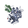
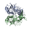
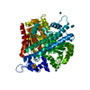
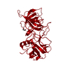
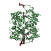
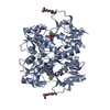
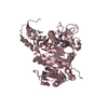
 Komagataeibacter europaeus (bacteria)
 X-RAY DIFFRACTION /
  Authors
 Citation
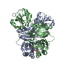
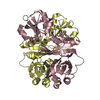
 Structure visualization
 Downloads & links
 Other downloads
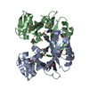


 PDBj
PDBj


 Assembly
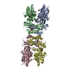

 Type: L-peptide linking / Mass: 165.189 Da / Num. of mol.: 4 / Source method: obtained synthetically / Formula: C9H11NO2
 Mass: 18.015 Da / Num. of mol.: 57 / Source method: isolated from a natural source / Formula: H2O
 Sample preparation
 / Beamline: P11 / Wavelength: 1.0332 Å
 Processing