 Movie
Movie Controller

[English] 日本語
 Yorodumi
- PDB-7al5: Crystal structure of the selenomethionine substituted hypothetica... -
+ Open data

- Basic information
| Entry | Database: PDB / ID: 7al5 | ||||||
|---|---|---|---|---|---|---|---|
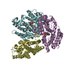
| Title | Crystal structure of the selenomethionine substituted hypothetical protein PA1622 from Pseudomonas aeruginosa PAO1 | ||||||
 Components Components | Probable hydrolase | ||||||
 Keywords Keywords | UNKNOWN FUNCTION / Esterase fold / hypothetical protein / Pseudomonas aeruginosa PAO1 / possible drug target / selenomethionine / Alpha/Beta hydrolase fold / Putative lipolytic enzyme | ||||||
| Function / homology | : / alpha/beta hydrolase fold / Alpha/beta hydrolase fold-1 / Alpha/Beta hydrolase fold / hydrolase activity / DI(HYDROXYETHYL)ETHER / TRIETHYLENE GLYCOL / Probable hydrolase Function and homology information Function and homology information | ||||||
| Biological species |   Pseudomonas aeruginosa (bacteria) Pseudomonas aeruginosa (bacteria) | ||||||
| Method |  X-RAY DIFFRACTION / SYNCHROTRON / SAD / Resolution: 2.42 Å X-RAY DIFFRACTION / SYNCHROTRON / SAD / Resolution: 2.42 Å | ||||||
 Authors Authors | Feiler, C.G. / Blankenfeldt, W. | ||||||
 Citation Citation | Journal: To Be Published Title: Crystal structure of the selenomethionine substituted hypothetical protein PA1622 from Pseudomonas aeruginosa PAO1 Authors: Feiler, C.G. / Blankenfeldt, W. | ||||||
| History |
|
- Structure visualization
| Structure viewer | Molecule: MolmilJmol/JSmol |
|---|
- Downloads & links
-Download
| PDBx/mmCIF format | 7al5.cif.gz | 425.1 KB | Display | PDBx/mmCIF format |
|---|---|---|---|---|
| PDB format | pdb7al5.ent.gz | 357.2 KB | Display | PDB format |
| PDBx/mmJSON format | 7al5.json.gz | Tree view | PDBx/mmJSON format | |
| Others |  Other downloads Other downloads |
-Validation report
| Summary document | 7al5_validation.pdf.gz | 490 KB | Display | wwPDB validaton report |
|---|---|---|---|---|
| Full document | 7al5_full_validation.pdf.gz | 504.6 KB | Display | |
| Data in XML | 7al5_validation.xml.gz | 47 KB | Display | |
| Data in CIF | 7al5_validation.cif.gz | 67.1 KB | Display | |
| Arichive directory | https://data.pdbj.org/pub/pdb/validation_reports/al/7al5ftp://data.pdbj.org/pub/pdb/validation_reports/al/7al5 | HTTPS FTP |
-Related structure data
| Similar structure data |
|---|


-Links
 PDBj
PDBj


- Assembly
| Deposited unit | 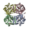
| ||||||||
|---|---|---|---|---|---|---|---|---|---|
| 1 |
| ||||||||
| Unit cell |
|
-Components
-Protein , 1 types, 4 molecules ABCD
| #1: Protein | Mass: 31662.555 Da / Num. of mol.: 4 Source method: isolated from a genetically manipulated source Source: (gene. exp.) Pseudomonas aeruginosa (strain ATCC 15692 / DSM 22644 / CIP 104116 / JCM 14847 / LMG 12228 / 1C / PRS 101 / PAO1) (bacteria)Strain: ATCC 15692 / DSM 22644 / CIP 104116 / JCM 14847 / LMG 12228 / 1C / PRS 101 / PAO1 Gene: PA1622 / Plasmid: P10$ / Production host: |
|---|
-Non-polymers , 5 types, 572 molecules 


| #2: Chemical |  Mass: 106.120 Da / Num. of mol.: 2 / Source method: obtained synthetically / Formula: C4H10O3 Mass: 106.120 Da / Num. of mol.: 2 / Source method: obtained synthetically / Formula: C4H10O3#3: Chemical | ChemComp-PGE / |  Mass: 150.173 Da / Num. of mol.: 1 / Source method: obtained synthetically / Formula: C6H14O4 Mass: 150.173 Da / Num. of mol.: 1 / Source method: obtained synthetically / Formula: C6H14O4#4: Chemical | ChemComp-EDO / |  Mass: 62.068 Da / Num. of mol.: 1 / Source method: obtained synthetically / Formula: C2H6O2 Mass: 62.068 Da / Num. of mol.: 1 / Source method: obtained synthetically / Formula: C2H6O2#5: Chemical | ChemComp-GOL / |  Mass: 92.094 Da / Num. of mol.: 1 / Source method: obtained synthetically / Formula: C3H8O3 Mass: 92.094 Da / Num. of mol.: 1 / Source method: obtained synthetically / Formula: C3H8O3#6: Water | ChemComp-HOH / | Mass: 18.015 Da / Num. of mol.: 567 / Source method: isolated from a natural source / Formula: H2O |
|---|
-Details
| Has ligand of interest | N |
|---|---|
| Has protein modification | Y |
-Experimental details
-Experiment
| Experiment | Method: X-RAY DIFFRACTION / Number of used crystals: 1 |
|---|
- Sample preparation
| Crystal | Density Matthews: 3.17 Å3/Da / Density % sol: 61.25 % |
|---|---|
| Crystal grow | Temperature: 293 K / Method: vapor diffusion, hanging drop / pH: 7 / Details: 0.1M HEPES pH7 5% PEG 6000 / PH range: 6.8 - 7.2 |
-Data collection
| Diffraction | Mean temperature: 100 K / Serial crystal experiment: N |
|---|---|
| Diffraction source | Source: SYNCHROTRON / Site: BESSY  / Beamline: 14.1 / Wavelength: 0.976 Å / Beamline: 14.1 / Wavelength: 0.976 Å |
| Detector | Type: MARMOSAIC 225 mm CCD / Detector: CCD / Date: Nov 14, 2012 |
| Radiation | Monochromator: Si111 / Protocol: SINGLE WAVELENGTH / Monochromatic (M) / Laue (L): M / Scattering type: x-ray |
| Radiation wavelength | Wavelength: 0.976 Å / Relative weight: 1 |
| Reflection | Resolution: 2.42→19.93 Å / Num. obs: 59611 / % possible obs: 99.6 % / Redundancy: 22.9 % / CC1/2: 0.988 / Rmerge(I) obs: 0.43 / Rpim(I) all: 0.094 / Rrim(I) all: 0.45 / Net I/σ(I): 10.2 |
| Reflection shell | Resolution: 2.42→2.48 Å / Rmerge(I) obs: 2.555 / Mean I/σ(I) obs: 1.3 / Num. unique obs: 4580 / CC1/2: 0.4 / Rpim(I) all: 0.633 / Rrim(I) all: 2.711 / Χ2: 1 |
- Processing
| Software |
| ||||||||||||||||||||||||||||||||||||||||||||||||||||||||||||||||||||||||||||||||||||||||||||||||||||||||||||||||||||||||||||||||||||||||||||||||||||||||||
|---|---|---|---|---|---|---|---|---|---|---|---|---|---|---|---|---|---|---|---|---|---|---|---|---|---|---|---|---|---|---|---|---|---|---|---|---|---|---|---|---|---|---|---|---|---|---|---|---|---|---|---|---|---|---|---|---|---|---|---|---|---|---|---|---|---|---|---|---|---|---|---|---|---|---|---|---|---|---|---|---|---|---|---|---|---|---|---|---|---|---|---|---|---|---|---|---|---|---|---|---|---|---|---|---|---|---|---|---|---|---|---|---|---|---|---|---|---|---|---|---|---|---|---|---|---|---|---|---|---|---|---|---|---|---|---|---|---|---|---|---|---|---|---|---|---|---|---|---|---|---|---|---|---|---|---|
| Refinement | Method to determine structure: SAD / Resolution: 2.42→19.93 Å / SU ML: 0.32 / Cross valid method: FREE R-VALUE / σ(F): 1.33 / Phase error: 23.9 / Stereochemistry target values: ML
| ||||||||||||||||||||||||||||||||||||||||||||||||||||||||||||||||||||||||||||||||||||||||||||||||||||||||||||||||||||||||||||||||||||||||||||||||||||||||||
| Solvent computation | Shrinkage radii: 0.9 Å / VDW probe radii: 1.11 Å / Solvent model: FLAT BULK SOLVENT MODEL | ||||||||||||||||||||||||||||||||||||||||||||||||||||||||||||||||||||||||||||||||||||||||||||||||||||||||||||||||||||||||||||||||||||||||||||||||||||||||||
| Refinement step | Cycle: LAST / Resolution: 2.42→19.93 Å
| ||||||||||||||||||||||||||||||||||||||||||||||||||||||||||||||||||||||||||||||||||||||||||||||||||||||||||||||||||||||||||||||||||||||||||||||||||||||||||
| Refine LS restraints |
| ||||||||||||||||||||||||||||||||||||||||||||||||||||||||||||||||||||||||||||||||||||||||||||||||||||||||||||||||||||||||||||||||||||||||||||||||||||||||||
| LS refinement shell |
|
