 Movie
Movie Controller

[English] 日本語
 Yorodumi
- PDB-3juk: The Crystal Structure of UDP-glucose pyrophosphorylase complexed ... -
+ Open data

- Basic information
| Entry | Database: PDB / ID: 3juk | ||||||
|---|---|---|---|---|---|---|---|
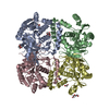
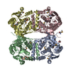
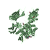
| Title | The Crystal Structure of UDP-glucose pyrophosphorylase complexed with UDP-glucose | ||||||
 Components Components | UDP-glucose pyrophosphorylase (GalU) | ||||||
 Keywords Keywords | TRANSFERASE / UDP-glucose pyrophosphorylase / Helicobacter pylori | ||||||
| Function / homology |  Function and homology information Function and homology informationUTP-glucose-1-phosphate uridylyltransferase / UTP:glucose-1-phosphate uridylyltransferase activity / UDP-alpha-D-glucose metabolic process / lipopolysaccharide core region biosynthetic process / metal ion binding / cytosol Similarity search - Function | ||||||
| Biological species |   Helicobacter pylori (bacteria) Helicobacter pylori (bacteria) | ||||||
| Method |  X-RAY DIFFRACTION / SYNCHROTRON / MOLECULAR REPLACEMENT / Resolution: 2.3 Å X-RAY DIFFRACTION / SYNCHROTRON / MOLECULAR REPLACEMENT / Resolution: 2.3 Å | ||||||
 Authors Authors | Kim, H. / Kim, K.K. | ||||||
 Citation Citation | Journal: Mol.Cells / Year: 2010 Title: Structural basis for the reaction mechanism of UDP-glucose pyrophosphorylase Authors: Kim, H. / Choi, J. / Kim, T. / Lokanath, N.K. / Ha, S.C. / Suh, S.W. / Hwang, H.-Y. / Kim, K.K. | ||||||
| History |
|
- Structure visualization
| Structure viewer | Molecule: MolmilJmol/JSmol |
|---|
- Downloads & links
-Download
| PDBx/mmCIF format | 3juk.cif.gz | 238.3 KB | Display | PDBx/mmCIF format |
|---|---|---|---|---|
| PDB format | pdb3juk.ent.gz | 191.1 KB | Display | PDB format |
| PDBx/mmJSON format | 3juk.json.gz | Tree view | PDBx/mmJSON format | |
| Others |  Other downloads Other downloads |
-Validation report
| Arichive directory | https://data.pdbj.org/pub/pdb/validation_reports/ju/3jukftp://data.pdbj.org/pub/pdb/validation_reports/ju/3juk | HTTPS FTP |
|---|
-Related structure data
| Related structure data | 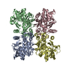 3jujSC S: Starting model for refinement C: citing same article ( |
|---|---|
| Similar structure data |

-Links
 PDBj
PDBj

- Assembly
| Deposited unit | 
| ||||||||
|---|---|---|---|---|---|---|---|---|---|
| 1 |
| ||||||||
| Unit cell |
|
-Components
| #1: Protein | Mass: 32086.932 Da / Num. of mol.: 4 Source method: isolated from a genetically manipulated source Source: (gene. exp.) Helicobacter pylori (bacteria) / Strain: 26695 / Gene: HP_0646 / Plasmid: pET-21a / Production host: References: UniProt: O25363, UTP-glucose-1-phosphate uridylyltransferase #2: Chemical | ChemComp-UPG /   Mass: 566.302 Da / Num. of mol.: 4 / Source method: obtained synthetically / Formula: C15H24N2O17P2 Mass: 566.302 Da / Num. of mol.: 4 / Source method: obtained synthetically / Formula: C15H24N2O17P2#3: Chemical | ChemComp-MG /   Mass: 24.305 Da / Num. of mol.: 12 / Source method: obtained synthetically / Formula: Mg Mass: 24.305 Da / Num. of mol.: 12 / Source method: obtained synthetically / Formula: Mg#4: Water | ChemComp-HOH / |  Mass: 18.015 Da / Num. of mol.: 736 / Source method: isolated from a natural source / Formula: H2O Mass: 18.015 Da / Num. of mol.: 736 / Source method: isolated from a natural source / Formula: H2O |
|---|
-Experimental details
-Experiment
| Experiment | Method: X-RAY DIFFRACTION / Number of used crystals: 1 |
|---|
- Sample preparation
| Crystal | Density Matthews: 2.43 Å3/Da / Density % sol: 49.45 % |
|---|---|
| Crystal grow | Temperature: 295 K / Method: vapor diffusion, hanging drop / pH: 7.5 Details: 0.1M HEPES-Na pH 7.5, 2% PEG 400, 1.5M ammonium sulfate, 10mM UDP-Glucose, 10 mM MgCl2 , VAPOR DIFFUSION, HANGING DROP, temperature 295K |
-Data collection
| Diffraction | Mean temperature: 100 K |
|---|---|
| Diffraction source | Source: SYNCHROTRON / Site: PAL/PLS  / Beamline: 4A / Wavelength: 1 Å / Beamline: 4A / Wavelength: 1 Å |
| Detector | Type: ADSC QUANTUM 210 / Detector: CCD / Date: May 29, 2007 |
| Radiation | Protocol: SINGLE WAVELENGTH / Monochromatic (M) / Laue (L): M / Scattering type: x-ray |
| Radiation wavelength | Wavelength: 1 Å / Relative weight: 1 |
| Reflection | Resolution: 2.3→50 Å / Num. all: 54678 / Num. obs: 54022 / % possible obs: 98.8 % / Redundancy: 3.1 % / Rsym value: 0.106 |
| Reflection shell | Resolution: 2.3→2.38 Å / Redundancy: 3.1 % / Num. unique all: 5102 / Rsym value: 0.368 / % possible all: 99.8 |
- Processing
| Software |
| ||||||||||||||||||||||||
|---|---|---|---|---|---|---|---|---|---|---|---|---|---|---|---|---|---|---|---|---|---|---|---|---|---|
| Refinement | Method to determine structure: MOLECULAR REPLACEMENT Starting model: PDB ENTRY 3JUJ Resolution: 2.3→50 Å / Occupancy max: 1 / Occupancy min: 1 / Cross valid method: THROUGHOUT / σ(F): 0
| ||||||||||||||||||||||||
| Solvent computation | Bsol: 87.582 Å2 | ||||||||||||||||||||||||
| Displacement parameters | Biso max: 73 Å2 / Biso mean: 25.749 Å2 / Biso min: 4.72 Å2
| ||||||||||||||||||||||||
| Refinement step | Cycle: LAST / Resolution: 2.3→50 Å
| ||||||||||||||||||||||||
| Refine LS restraints |
| ||||||||||||||||||||||||
| Xplor file |
|
