 Movie
Movie Controller


+ Open data

- Basic information
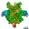
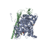
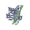
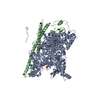
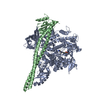
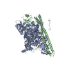
| Entry | Database: PDB / ID: 7agg | |||||||||||||||||||||||||||
|---|---|---|---|---|---|---|---|---|---|---|---|---|---|---|---|---|---|---|---|---|---|---|---|---|---|---|---|---|
| Title | HAd7 knob in complex with 2 EC2-EC3 modules of DSG-2 | |||||||||||||||||||||||||||
 Components Components |
| |||||||||||||||||||||||||||
 Keywords Keywords | VIRAL PROTEIN / adenovirus / cell receptor / complex | |||||||||||||||||||||||||||
| Function / homology |  Function and homology information Function and homology informationPurkinje myocyte development / positive regulation of protein localization to cell-cell junction / bundle of His cell-Purkinje myocyte adhesion involved in cell communication / cell adhesive protein binding involved in bundle of His cell-Purkinje myocyte communication / desmosome organization / negative regulation of endothelial cell differentiation / Keratinization / negative regulation of inflammatory response to wounding / desmosome / mesenchymal to epithelial transition ...Purkinje myocyte development / positive regulation of protein localization to cell-cell junction / bundle of His cell-Purkinje myocyte adhesion involved in cell communication / cell adhesive protein binding involved in bundle of His cell-Purkinje myocyte communication / desmosome organization / negative regulation of endothelial cell differentiation / Keratinization / negative regulation of inflammatory response to wounding / desmosome / mesenchymal to epithelial transition / Formation of the cornified envelope / cornified envelope / Apoptotic cleavage of cell adhesion proteins / regulation of ventricular cardiac muscle cell action potential / adhesion receptor-mediated virion attachment to host cell / negative regulation of epithelial to mesenchymal transition / positive regulation of sprouting angiogenesis / homophilic cell-cell adhesion / positive regulation of stem cell population maintenance / regulation of heart rate by cardiac conduction / RHOG GTPase cycle / intercalated disc / lateral plasma membrane / RAC3 GTPase cycle / RAC2 GTPase cycle / maternal process involved in female pregnancy / response to progesterone / cell adhesion molecule binding / positive regulation of cell adhesion / stem cell proliferation / cell-cell adhesion / cell-cell junction / viral capsid / cell junction / cell adhesion / apical plasma membrane / calcium ion binding / symbiont entry into host cell / negative regulation of apoptotic process / host cell nucleus / cell surface / extracellular exosome / plasma membrane / cytoplasm Similarity search - Function | |||||||||||||||||||||||||||
| Biological species |  Human adenovirus B serotype 7 Human adenovirus B serotype 7 Homo sapiens (human) Homo sapiens (human) | |||||||||||||||||||||||||||
| Method | ELECTRON MICROSCOPY / single particle reconstruction / cryo EM / Resolution: 3.3 Å | |||||||||||||||||||||||||||
 Authors Authors | Effantin, G. / Vassal-Stermann, E. / Fender, P. | |||||||||||||||||||||||||||
| Funding support |  France, 1items France, 1items
| |||||||||||||||||||||||||||
 Citation Citation | Journal: Viruses / Year: 2020 Title: Binding Mechanism Elucidation of the Acute Respiratory Disease Causing Agent Adenovirus of Serotype 7 to Desmoglein-2. Authors: Marc-André Hograindleur / Gregory Effantin / Daphna Fenel / Caroline Mas / André Lieber / Guy Schoehn / Pascal Fender / Emilie Vassal-Stermann /  Abstract: The study of viruses causing acute respiratory distress syndromes (ARDS) is more essential than ever at a time when a virus can create a global pandemic in a matter of weeks. Among human ...The study of viruses causing acute respiratory distress syndromes (ARDS) is more essential than ever at a time when a virus can create a global pandemic in a matter of weeks. Among human adenoviruses, adenovirus of serotype 7 (HAdV7) is one of the most virulent serotypes. This virus regularly re-emerges in Asia and has just been the cause of several deaths in the United States. A critical step of the virus life cycle is the attachment of the knob domain of the fiber (HAd7K) to the cellular receptor desmoglein-2 (DSG2). Complexes between the fiber knob and two extracellular domains of DSG2 have been produced. Their characterization by biochemical and biophysical methods show that these two domains are sufficient for the interaction and that the trimeric HAd7K could accommodate up to three DSG2 receptor molecules. The cryo-electron microscopy (cryo-EM) structure of these complexes at 3.1 Å resolution confirmed the biochemical data, and allowed the identification of the critical amino acid residues for this interaction, which shows similarities with other DSG2 interacting adenoviruses, despite a low homology in the primary sequences. | |||||||||||||||||||||||||||
| History |
|
- Structure visualization
| Movie |
Movie viewer |
|---|---|
| Structure viewer | Molecule: MolmilJmol/JSmol |
- Downloads & links
-Download
| PDBx/mmCIF format | 7agg.cif.gz | 189.4 KB | Display | PDBx/mmCIF format |
|---|---|---|---|---|
| PDB format | pdb7agg.ent.gz | 150.3 KB | Display | PDB format |
| PDBx/mmJSON format | 7agg.json.gz | Tree view | PDBx/mmJSON format | |
| Others |  Other downloads Other downloads |
-Validation report
| Arichive directory | https://data.pdbj.org/pub/pdb/validation_reports/ag/7aggftp://data.pdbj.org/pub/pdb/validation_reports/ag/7agg | HTTPS FTP |
|---|
-Related structure data
| Related structure data | 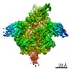 11779MC 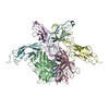 7agfC C: citing same article ( M: map data used to model this data |
|---|---|
| Similar structure data |


-Links
 PDBj
PDBj


- Assembly
| Deposited unit | 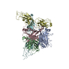
|
|---|---|
| 1 |
|
-Components
| #1: Protein | Mass: 23489.217 Da / Num. of mol.: 3 Source method: isolated from a genetically manipulated source Source: (gene. exp.) Human adenovirus B serotype 7 / Gene: L5 / Production host:  #2: Protein | Mass: 27126.275 Da / Num. of mol.: 2 Source method: isolated from a genetically manipulated source Source: (gene. exp.) Homo sapiens (human) / Gene: DSG2, CDHF5 / Production host: Homo sapiens (human) / References: UniProt: Q14126Has protein modification | N | |
|---|
-Experimental details
-Experiment
| Experiment | Method: ELECTRON MICROSCOPY |
|---|---|
| EM experiment | Aggregation state: PARTICLE / 3D reconstruction method: single particle reconstruction |
- Sample preparation
| Component |
| ||||||||||||||||||||||||
|---|---|---|---|---|---|---|---|---|---|---|---|---|---|---|---|---|---|---|---|---|---|---|---|---|---|
| Molecular weight | Value: 0.11 MDa / Experimental value: YES | ||||||||||||||||||||||||
| Source (natural) |
| ||||||||||||||||||||||||
| Source (recombinant) |
| ||||||||||||||||||||||||
| Buffer solution | pH: 8 | ||||||||||||||||||||||||
| Specimen | Conc.: 0.1 mg/ml / Embedding applied: NO / Shadowing applied: NO / Staining applied: NO / Vitrification applied: YES | ||||||||||||||||||||||||
| Vitrification | Cryogen name: ETHANE |
- Electron microscopy imaging
| Experimental equipment |  Model: Titan Krios / Image courtesy: FEI Company |
|---|---|
| Microscopy | Model: FEI TITAN KRIOS |
| Electron gun | Electron source:  FIELD EMISSION GUN / Accelerating voltage: 300 kV / Illumination mode: FLOOD BEAM FIELD EMISSION GUN / Accelerating voltage: 300 kV / Illumination mode: FLOOD BEAM |
| Electron lens | Mode: BRIGHT FIELD |
| Image recording | Electron dose: 40 e/Å2 / Film or detector model: GATAN K2 SUMMIT (4k x 4k) |
| EM imaging optics | Phase plate: VOLTA PHASE PLATE |
- Processing
| Software | Name: PHENIX / Version: 1.18.2_3874: / Classification: refinement | ||||||||||||||||||||||||
|---|---|---|---|---|---|---|---|---|---|---|---|---|---|---|---|---|---|---|---|---|---|---|---|---|---|
| EM software | Name: PHENIX / Category: model refinement | ||||||||||||||||||||||||
| CTF correction | Type: PHASE FLIPPING AND AMPLITUDE CORRECTION | ||||||||||||||||||||||||
| 3D reconstruction | Resolution: 3.3 Å / Resolution method: FSC 0.143 CUT-OFF / Num. of particles: 97190 / Symmetry type: POINT | ||||||||||||||||||||||||
| Refine LS restraints |
|
