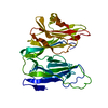
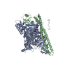
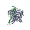
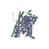
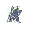
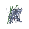
Entry Database : PDB / ID : 6nctTitle Structure of p110alpha/niSH2 - vector data collection Phosphatidylinositol 3-kinase regulatory subunit alpha Phosphatidylinositol 4,5-bisphosphate 3-kinase catalytic subunit alpha isoform Keywords / / / / / / Function / homology Function Domain/homology Component
/ / / / / / / / / / / / / / / / / / / / / / / / / / / / / / / / / / / / / / / / / / / / / / / / / / / / / / / / / / / / / / / / / / / / / / / / / / / / / / / / / / / / / / / / / / / / / / / / / / / / / / / / / / / / / / / / / / / / / / / / / / / / / / / / / / / / / / / / / / / / / / / / / / / / / / / / / / / / / / / / / / / / / / / / / / / / / / / / / / / / Biological species Homo sapiens (human)Method / / / / Resolution : 3.35 Å Authors Miller, M.S. / Maheshwari, S. / Amzel, L.M. / Gabelli, S.B. Funding support Organization Grant number Country National Institutes of Health/National Cancer Institute (NIH/NCI) CA062924
Journal : Molecules / Year : 2019Title : Getting the Most Out of Your Crystals: Data Collection at the New High-Flux, Microfocus MX Beamlines at NSLS-II.Authors : Miller, M.S. / Maheshwari, S. / Shi, W. / Gao, Y. / Chu, N. / Soares, A.S. / Cole, P.A. / Amzel, L.M. / Fuchs, M.R. / Jakoncic, J. / Gabelli, S.B. History Deposition Dec 12, 2018 Deposition site / Processing site Revision 1.0 Feb 6, 2019 Provider / Type Revision 1.1 Nov 13, 2019 Group / Category / citation_authorItem _citation.journal_volume / _citation.page_first ... _citation.journal_volume / _citation.page_first / _citation.pdbx_database_id_PubMed / _citation.title / _citation_author.identifier_ORCID Revision 1.2 Dec 4, 2019 Group / Category / Item Revision 1.3 Oct 11, 2023 Group / Database references / Refinement descriptionCategory chem_comp_atom / chem_comp_bond ... chem_comp_atom / chem_comp_bond / database_2 / pdbx_initial_refinement_model Item / _database_2.pdbx_database_accession
Show all Show less
 Movie
Movie Controller


 Open data
 Basic information
 Components
 Keywords
 Function and homology information
 Homo sapiens (human)
 X-RAY DIFFRACTION /
  Authors
 United States, 1items
  Citation
 Structure visualization
 Downloads & links
 Other downloads


 PDBj
PDBj


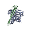
 Assembly
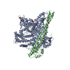

 Spodoptera frugiperda (fall armyworm)
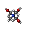
 Mass: 122.143 Da / Num. of mol.: 2 / Source method: obtained synthetically / Formula: C4H12NO3
 Mass: 96.063 Da / Num. of mol.: 3 / Source method: obtained synthetically / Formula: SO4
 Sample preparation
 Processing