 Movie
Movie Controller


+ Open data

- Basic information
| Entry | Database: PDB / ID: 6nci | |||||||||
|---|---|---|---|---|---|---|---|---|---|---|
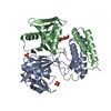
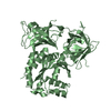
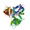
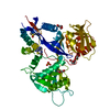
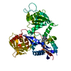
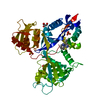
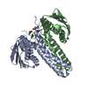
| Title | Crystal structure of CDP-Chase: Vector data collection | |||||||||
 Components Components | Phosphohydrolase (MutT/nudix family protein) | |||||||||
 Keywords Keywords | HYDROLASE / nudix / CDP-chase / CDP-choline hydrolase / ADP-ribose / vector data collection | |||||||||
| Function / homology |  Function and homology information Function and homology informationdihydroneopterin triphosphate pyrophosphohydrolase activity / dATP diphosphatase activity / folic acid biosynthetic process / tetrahydrofolate biosynthetic process Similarity search - Function | |||||||||
| Biological species |  | |||||||||
| Method |  X-RAY DIFFRACTION / SYNCHROTRON / MOLECULAR REPLACEMENT / molecular replacement / Resolution: 2.08 Å X-RAY DIFFRACTION / SYNCHROTRON / MOLECULAR REPLACEMENT / molecular replacement / Resolution: 2.08 Å | |||||||||
 Authors Authors | Miller, M.S. / Shi, W. / Gabelli, S.B. | |||||||||
| Funding support |  United States, 1items United States, 1items
| |||||||||
 Citation Citation | Journal: Molecules / Year: 2019 Title: Getting the Most Out of Your Crystals: Data Collection at the New High-Flux, Microfocus MX Beamlines at NSLS-II. Authors: Miller, M.S. / Maheshwari, S. / Shi, W. / Gao, Y. / Chu, N. / Soares, A.S. / Cole, P.A. / Amzel, L.M. / Fuchs, M.R. / Jakoncic, J. / Gabelli, S.B. | |||||||||
| History |
|
- Structure visualization
| Structure viewer | Molecule: MolmilJmol/JSmol |
|---|
- Downloads & links
-Download
| PDBx/mmCIF format | 6nci.cif.gz | 103.4 KB | Display | PDBx/mmCIF format |
|---|---|---|---|---|
| PDB format | pdb6nci.ent.gz | 77.2 KB | Display | PDB format |
| PDBx/mmJSON format | 6nci.json.gz | Tree view | PDBx/mmJSON format | |
| Others |  Other downloads Other downloads |
-Validation report
| Arichive directory | https://data.pdbj.org/pub/pdb/validation_reports/nc/6nciftp://data.pdbj.org/pub/pdb/validation_reports/nc/6nci | HTTPS FTP |
|---|
-Related structure data
| Related structure data |  6nchC 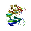 6nckC  6nctC  3q1pS S: Starting model for refinement C: citing same article ( |
|---|---|
| Similar structure data |


-Links
 PDBj
PDBj

- Assembly
| Deposited unit | 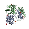
| ||||||||
|---|---|---|---|---|---|---|---|---|---|
| 1 |
| ||||||||
| Unit cell |
|
-Components
-Protein / Sugars , 2 types, 3 molecules AB
| #1: Protein | Mass: 23718.875 Da / Num. of mol.: 2 Source method: isolated from a genetically manipulated source Source: (gene. exp.) Strain: ATCC 14579 / DSM 31 / JCM 2152 / NBRC 15305 / NCIMB 9373 / NRRL B-3711 Gene: BC_2032 / Production host: #5: Sugar | ChemComp-RB5 / |  Type: D-saccharide / Mass: 150.130 Da / Num. of mol.: 1 Type: D-saccharide / Mass: 150.130 Da / Num. of mol.: 1Source method: isolated from a genetically manipulated source Formula: C5H10O5 / Feature type: SUBJECT OF INVESTIGATION |
|---|
-Non-polymers , 4 types, 263 molecules 


| #2: Chemical | ChemComp-SO4 /  Mass: 96.063 Da / Num. of mol.: 6 / Source method: obtained synthetically / Formula: SO4 Mass: 96.063 Da / Num. of mol.: 6 / Source method: obtained synthetically / Formula: SO4#3: Chemical | ChemComp-PO4 / |  Mass: 94.971 Da / Num. of mol.: 1 / Source method: obtained synthetically / Formula: PO4 / Feature type: SUBJECT OF INVESTIGATION Mass: 94.971 Da / Num. of mol.: 1 / Source method: obtained synthetically / Formula: PO4 / Feature type: SUBJECT OF INVESTIGATION#4: Chemical | ChemComp-PEG / |  Mass: 106.120 Da / Num. of mol.: 1 / Source method: obtained synthetically / Formula: C4H10O3 Mass: 106.120 Da / Num. of mol.: 1 / Source method: obtained synthetically / Formula: C4H10O3#6: Water | ChemComp-HOH / | Mass: 18.015 Da / Num. of mol.: 255 / Source method: isolated from a natural source / Formula: H2O |
|---|
-Experimental details
-Experiment
| Experiment | Method: X-RAY DIFFRACTION / Number of used crystals: 1 |
|---|
- Sample preparation
| Crystal | Density Matthews: 2.43 Å3/Da / Density % sol: 49.4 % |
|---|---|
| Crystal grow | Temperature: 298 K / Method: vapor diffusion, hanging drop / pH: 8.5 Details: 0.1 M Tris-HCl pH 8.5, 0.2 to 0.3 M Lithium sulfate, 26 to 29% PEG-4000 |
-Data collection
| Diffraction | Mean temperature: 100 K / Serial crystal experiment: N |
|---|---|
| Diffraction source | Source: SYNCHROTRON / Site: NSLS-II / Beamline: 17-ID-2 / Wavelength: 0.9793 Å |
| Detector | Type: DECTRIS EIGER X 16M / Detector: PIXEL / Date: Mar 29, 2018 |
| Radiation | Protocol: SINGLE WAVELENGTH / Monochromatic (M) / Laue (L): M / Scattering type: x-ray |
| Radiation wavelength | Wavelength: 0.9793 Å / Relative weight: 1 |
| Reflection | Resolution: 2.08→29.47 Å / Num. obs: 28297 / % possible obs: 98.8 % / Redundancy: 13 % / CC1/2: 0.998 / Rmerge(I) obs: 0.136 / Rpim(I) all: 0.039 / Rrim(I) all: 0.142 / Net I/σ(I): 14.4 |
| Reflection shell | Resolution: 2.08→2.13 Å / Redundancy: 11.2 % / Rmerge(I) obs: 0.942 / Num. unique obs: 1769 / CC1/2: 0.818 / Rpim(I) all: 0.28 / Rrim(I) all: 0.985 / % possible all: 84.5 |
-Phasing
| Phasing | Method: molecular replacement | ||||||
|---|---|---|---|---|---|---|---|
| Phasing MR | R rigid body: 0.503
|
- Processing
| Software |
| ||||||||||||||||||||||||||||||||||||||||||||||||||||||||||||
|---|---|---|---|---|---|---|---|---|---|---|---|---|---|---|---|---|---|---|---|---|---|---|---|---|---|---|---|---|---|---|---|---|---|---|---|---|---|---|---|---|---|---|---|---|---|---|---|---|---|---|---|---|---|---|---|---|---|---|---|---|---|
| Refinement | Method to determine structure: MOLECULAR REPLACEMENT Starting model: 3Q1P Resolution: 2.08→29.47 Å / Cor.coef. Fo:Fc: 0.962 / Cor.coef. Fo:Fc free: 0.933 / SU B: 4.656 / SU ML: 0.125 / SU R Cruickshank DPI: 0.1962 / Cross valid method: THROUGHOUT / σ(F): 0 / ESU R: 0.196 / ESU R Free: 0.173 Details: HYDROGENS HAVE BEEN ADDED IN THE RIDING POSITIONS U VALUES : REFINED INDIVIDUALLY
| ||||||||||||||||||||||||||||||||||||||||||||||||||||||||||||
| Solvent computation | Ion probe radii: 0.8 Å / Shrinkage radii: 0.8 Å / VDW probe radii: 1.2 Å | ||||||||||||||||||||||||||||||||||||||||||||||||||||||||||||
| Displacement parameters | Biso max: 112.64 Å2 / Biso mean: 33.807 Å2 / Biso min: 16.23 Å2
| ||||||||||||||||||||||||||||||||||||||||||||||||||||||||||||
| Refinement step | Cycle: final / Resolution: 2.08→29.47 Å
| ||||||||||||||||||||||||||||||||||||||||||||||||||||||||||||
| Refine LS restraints |
| ||||||||||||||||||||||||||||||||||||||||||||||||||||||||||||
| LS refinement shell | Resolution: 2.078→2.132 Å / Rfactor Rfree error: 0 / Total num. of bins used: 20
|
