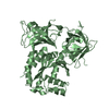
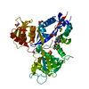
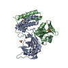
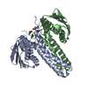
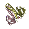
- PDB-4fmt: Crystal structure of a ChpT protein (CC_3470) from Caulobacter cr... -
+
Open data
ID or keywords:
Loading...
-
Basic information
Entry
Database: PDB / ID: 4fmt
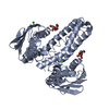
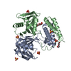
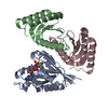
Title
Crystal structure of a ChpT protein (CC_3470) from Caulobacter crescentus CB15 at 2.30 A resolution
Components
ChpT protein
Keywords
TRANSFERASE / a phosphotransfer protein / a two-component signaling pathway / Structural Genomics / Joint Center for Structural Genomics / JCSG / Protein Structure Initiative / PSI-BIOLOGY
Mass: 18.015 Da / Num. of mol.: 311 / Source method: isolated from a natural source / Formula: H2O
Sequence details
THE CONSTRUCT WAS EXPRESSED WITH AN N-TERMINAL PURIFICATION TAG MGSSHHHHHHSSGLVPRGSH. THE TAG WAS ...THE CONSTRUCT WAS EXPRESSED WITH AN N-TERMINAL PURIFICATION TAG MGSSHHHHHHSSGLVPRGSH. THE TAG WAS REMOVED WITH THROMBIN LEAVING ONLY (-2)-GLY-SER-HIS-(0) FOLLOWED BY THE FULL LENGTH TARGET SEQUENCE (1-225). THE START CODON FOR UNIPROT ENTRY A6H916 WAS MIS-ANNOTATED (SEE PMID 21878915). THE CLONED CONSTRUCT STARTS FROM THE CORRECT START CODON THAT CORRESPONDS TO MET-29 OF UNIPROT ENTRY A6H916 (VERSION 1 OF THE SEQUENCE).
-
Experimental details
-
Experiment
Experiment
Method: X-RAY DIFFRACTION / Number of used crystals: 2
20% (w/v) PEG-8000, 0.1M MES pH 6.0, 0.2M Calcium acetate, final pH 6.2, VAPOR DIFFUSION,HANGING DROP, temperature 295.5K
295.5
2
vapor diffusion, hanging drop
6.2
20% (w/v) PEG-8000, 0.1M MES pH 6.0, 0.2M Calcium acetate, final pH 6.2, VAPOR DIFFUSION,HANGING DROP, temperature 295.5K
Experiment crystal cryo treatment
Crystal-ID
Cooling details
Final solution details
Soaking details
1
Directimmersioninliquidnitrogen
30% (w/v) PEG-8000 in precipitant
2
Directimmersioninliquidnitrogen
30% (w/v) PEG-8000, 0.01 M potassium dicyanoaurate (I) in precipitant
First the PEG-8000 was increased to 30% (w/v) PEG-8000 in precipitant. Potassium dicyanoaurate (I) was then added to a final concentration of 0.010 M and after 190 minutes the crystal was harvested.
Resolution: 2.3→65.094 Å / Num. obs: 52631 / % possible obs: 97.2 % / Observed criterion σ(I): -3 / Redundancy: 3.45 % / Biso Wilson estimate: 51.445 Å2 / Rmerge(I) obs: 0.087 / Net I/σ(I): 10.67
Reflection shell
Diffraction-ID: 1,2
Resolution (Å)
Redundancy (%)
Rmerge(I) obs
Mean I/σ(I) obs
Num. measured obs
Num. unique obs
% possible all
2.3-2.36
3.43
0.912
1.64
13267
3869
97.6
2.36-2.42
3.4
0.77
2
12793
3771
97
2.42-2.49
3.3
0.697
2.2
11667
3579
94.3
2.49-2.57
3.5
0.484
3.4
12491
3570
98
2.57-2.66
3.6
0.405
4.4
12906
3554
99.4
2.66-2.75
3.6
0.31
5.4
12385
3431
99.4
2.75-2.85
3.6
0.269
6.1
11980
3321
99.2
2.85-2.97
3.6
0.198
7.8
11331
3166
99.2
2.97-3.1
3.5
0.161
9.2
10632
3040
98.7
3.1-3.25
3.3
0.123
10.7
8996
2755
94.4
3.25-3.43
3.6
0.087
14.5
9843
2759
98
3.43-3.64
3.5
0.068
17.5
9066
2572
97.7
3.64-3.89
3.4
0.057
19.3
8264
2403
96.7
3.89-4.2
3.4
0.05
20.9
7602
2252
96.6
4.2-4.6
3.2
0.042
23.6
6539
2023
94.4
4.6-5.14
3.3
0.038
24
5801
1784
92.5
5.14-5.94
3.5
0.041
23.5
5913
1697
97.9
5.94-7.27
3.4
0.036
24.2
4825
1411
96.5
7.27-10.29
3.1
0.027
27.7
3341
1066
94.9
10.29-65.09
3.5
0.029
30.7
2121
607
94.1
-
Phasing
Phasing
Method: MAD
-
Processing
Software
Name
Version
Classification
NB
MolProbity
3beta29
modelbuilding
PDB_EXTRACT
3.1
dataextraction
SHELX
phasing
SHARP
phasing
XSCALE
December6, 2010
datascaling
BUSTER-TNT
2.10.0
refinement
XDS
datareduction
SHELXD
phasing
BUSTER
2.10.0
refinement
Refinement
Method to determine structure: MAD / Resolution: 2.3→65.094 Å / Cor.coef. Fo:Fc: 0.9408 / Cor.coef. Fo:Fc free: 0.917 / Occupancy max: 1 / Occupancy min: 0.4 / Cross valid method: THROUGHOUT / σ(F): 0 Details: 1. ATOM RECORD CONTAINS SUM OF TLS AND RESIDUAL B FACTORS. ANISOU RECORD CONTAINS SUM OF TLS AND RESIDUAL U FACTORS. 2. THE MAD PHASES WERE USED AS RESTRAINTS DURING REFINEMENT EXCEPT FOR ...Details: 1. ATOM RECORD CONTAINS SUM OF TLS AND RESIDUAL B FACTORS. ANISOU RECORD CONTAINS SUM OF TLS AND RESIDUAL U FACTORS. 2. THE MAD PHASES WERE USED AS RESTRAINTS DURING REFINEMENT EXCEPT FOR THE FINAL REFINEMENT CYCLE. 3. NCS RESTRAINTS WERE APPLIED USING BUSTER'S LSSR RESTRAINT REPRESENTATION (-AUTONCS). 4. NA IONS AND GLYCEROL MODELED ARE PRESENT IN PURIFICATION/CRYSTALLIZATION CONDITIONS.
In the structure databanks used in Yorodumi, some data are registered as the other names, "COVID-19 virus" and "2019-nCoV". Here are the details of the virus and the list of structure data.
Jan 31, 2019. EMDB accession codes are about to change! (news from PDBe EMDB page)
EMDB accession codes are about to change! (news from PDBe EMDB page)
The allocation of 4 digits for EMDB accession codes will soon come to an end. Whilst these codes will remain in use, new EMDB accession codes will include an additional digit and will expand incrementally as the available range of codes is exhausted. The current 4-digit format prefixed with “EMD-” (i.e. EMD-XXXX) will advance to a 5-digit format (i.e. EMD-XXXXX), and so on. It is currently estimated that the 4-digit codes will be depleted around Spring 2019, at which point the 5-digit format will come into force.
The EM Navigator/Yorodumi systems omit the EMD- prefix.
Related info.:Q: What is EMD? / ID/Accession-code notation in Yorodumi/EM Navigator
Yorodumi is a browser for structure data from EMDB, PDB, SASBDB, etc.
This page is also the successor to EM Navigator detail page, and also detail information page/front-end page for Omokage search.
The word "yorodu" (or yorozu) is an old Japanese word meaning "ten thousand". "mi" (miru) is to see.
Related info.:EMDB / PDB / SASBDB / Comparison of 3 databanks / Yorodumi Search / Aug 31, 2016. New EM Navigator & Yorodumi / Yorodumi Papers / Jmol/JSmol / Function and homology information / Changes in new EM Navigator and Yorodumi
 Movie
Movie Controller

 Yorodumi
 Open data
 Basic information
 Components
 Keywords
 Function and homology information
 Caulobacter crescentus (bacteria)
 X-RAY DIFFRACTION /
  Authors
 Citation
 Structure visualization
 Downloads & links
 Other downloads
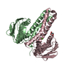


 PDBj
PDBj Assembly
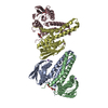

 Mass: 22.990 Da / Num. of mol.: 4 / Source method: obtained synthetically / Formula: Na
 Mass: 92.094 Da / Num. of mol.: 4 / Source method: obtained synthetically / Formula: C3H8O3
 Mass: 18.015 Da / Num. of mol.: 311 / Source method: isolated from a natural source / Formula: H2O
 Sample preparation
 Processing