 Movie
Movie Controller


+ Open data

- Basic information
| Entry | Database: PDB / ID: 7abf | |||||||||||||||||||||
|---|---|---|---|---|---|---|---|---|---|---|---|---|---|---|---|---|---|---|---|---|---|---|
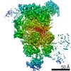
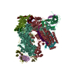
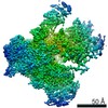
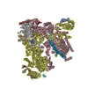
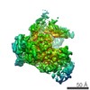
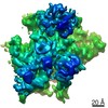
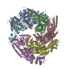
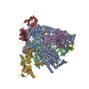
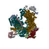
| Title | Human pre-Bact-1 spliceosome core structure | |||||||||||||||||||||
 Components Components |
| |||||||||||||||||||||
 Keywords Keywords | SPLICING / Complex / spliceosome / catalytic activation | |||||||||||||||||||||
| Function / homology |  Function and homology information Function and homology informationmicrofibril / regulation of retinoic acid receptor signaling pathway / regulation of vitamin D receptor signaling pathway / nuclear retinoic acid receptor binding / U2-type catalytic step 1 spliceosome / RNA splicing, via transesterification reactions / positive regulation of mRNA splicing, via spliceosome / positive regulation of vitamin D receptor signaling pathway / host-mediated activation of viral transcription / U2-type precatalytic spliceosome ...microfibril / regulation of retinoic acid receptor signaling pathway / regulation of vitamin D receptor signaling pathway / nuclear retinoic acid receptor binding / U2-type catalytic step 1 spliceosome / RNA splicing, via transesterification reactions / positive regulation of mRNA splicing, via spliceosome / positive regulation of vitamin D receptor signaling pathway / host-mediated activation of viral transcription / U2-type precatalytic spliceosome / mRNA cis splicing, via spliceosome / Regulation of gene expression in late stage (branching morphogenesis) pancreatic bud precursor cells / RNA polymerase binding / Notch binding / RUNX3 regulates NOTCH signaling / U2-type catalytic step 2 spliceosome / U2-type spliceosomal complex / nuclear vitamin D receptor binding / NOTCH4 Intracellular Domain Regulates Transcription / transcription elongation factor activity / NOTCH3 Intracellular Domain Regulates Transcription / : / positive regulation of neurogenesis / K63-linked polyubiquitin modification-dependent protein binding / nuclear androgen receptor binding / precatalytic spliceosome / Notch-HLH transcription pathway / WW domain binding / Formation of paraxial mesoderm / ubiquitin-like protein conjugating enzyme binding / positive regulation of transforming growth factor beta receptor signaling pathway / SMAD binding / mRNA Splicing - Minor Pathway / spliceosomal tri-snRNP complex assembly / Prp19 complex / negative regulation of transcription elongation by RNA polymerase II / U5 snRNP / U5 snRNA binding / intrinsic apoptotic signaling pathway in response to DNA damage by p53 class mediator / pre-mRNA intronic binding / U2 snRNA binding / U6 snRNA binding / protein localization to nucleus / Cajal body / U1 snRNA binding / positive regulation of G1/S transition of mitotic cell cycle / U4/U6 x U5 tri-snRNP complex / retinoic acid receptor signaling pathway / cellular response to retinoic acid / catalytic step 2 spliceosome / mRNA Splicing - Major Pathway / RNA splicing / acrosomal vesicle / nuclear receptor binding / spliceosomal complex / transcription coregulator activity / response to cocaine / positive regulation of transcription elongation by RNA polymerase II / sperm end piece / Downregulation of SMAD2/3:SMAD4 transcriptional activity / mRNA splicing, via spliceosome / NOTCH1 Intracellular Domain Regulates Transcription / Pre-NOTCH Transcription and Translation / cellular response to tumor necrosis factor / cellular response to xenobiotic stimulus / Constitutive Signaling by NOTCH1 PEST Domain Mutants / Constitutive Signaling by NOTCH1 HD+PEST Domain Mutants / fibrillar center / nuclear matrix / mRNA processing / protein tag activity / rRNA processing / transcription corepressor activity / single-stranded DNA binding / sperm principal piece / cellular response to lipopolysaccharide / microtubule cytoskeleton / nuclear membrane / RNA polymerase II-specific DNA-binding transcription factor binding / transcription coactivator activity / nuclear speck / nuclear body / negative regulation of DNA-templated transcription / GTPase activity / regulation of transcription by RNA polymerase II / centrosome / chromatin / GTP binding / enzyme binding / negative regulation of transcription by RNA polymerase II / positive regulation of transcription by RNA polymerase II / mitochondrion / DNA binding / RNA binding / zinc ion binding / nucleoplasm / membrane / identical protein binding / nucleus / cytoplasm Similarity search - Function | |||||||||||||||||||||
| Biological species |  Homo sapiens (human) Homo sapiens (human)synthetic construct (others) | |||||||||||||||||||||
| Method | ELECTRON MICROSCOPY / single particle reconstruction / cryo EM / Resolution: 3.9 Å | |||||||||||||||||||||
 Authors Authors | Townsend, C. / Kastner, B. / Leelaram, M.N. / Bertram, K. / Stark, H. / Luehrmann, R. | |||||||||||||||||||||
| Funding support |  Germany, 1items Germany, 1items
| |||||||||||||||||||||
 Citation Citation | Journal: Science / Year: 2020 Title: Mechanism of protein-guided folding of the active site U2/U6 RNA during spliceosome activation. Authors: Cole Townsend / Majety N Leelaram / Dmitry E Agafonov / Olexandr Dybkov / Cindy L Will / Karl Bertram / Henning Urlaub / Berthold Kastner / Holger Stark / Reinhard Lührmann / Abstract: Spliceosome activation involves extensive protein and RNA rearrangements that lead to formation of a catalytically active U2/U6 RNA structure. At present, little is known about the assembly pathway ...Spliceosome activation involves extensive protein and RNA rearrangements that lead to formation of a catalytically active U2/U6 RNA structure. At present, little is known about the assembly pathway of the latter and the mechanism whereby proteins aid its proper folding. Here, we report the cryo-electron microscopy structures of two human, activated spliceosome precursors (that is, pre-B complexes) at core resolutions of 3.9 and 4.2 angstroms. These structures elucidate the order of the numerous protein exchanges that occur during activation, the mutually exclusive interactions that ensure the correct order of ribonucleoprotein rearrangements needed to form the U2/U6 catalytic RNA, and the stepwise folding pathway of the latter. Structural comparisons with mature B complexes reveal the molecular mechanism whereby a conformational change in the scaffold protein PRP8 facilitates final three-dimensional folding of the U2/U6 catalytic RNA. | |||||||||||||||||||||
| History |
|
- Structure visualization
| Movie |
Movie viewer |
|---|---|
| Structure viewer | Molecule: MolmilJmol/JSmol |
- Downloads & links
-Download
| PDBx/mmCIF format | 7abf.cif.gz | 757.8 KB | Display | PDBx/mmCIF format |
|---|---|---|---|---|
| PDB format | pdb7abf.ent.gz | Display | PDB format | |
| PDBx/mmJSON format | 7abf.json.gz | Tree view | PDBx/mmJSON format | |
| Others |  Other downloads Other downloads |
-Validation report
| Arichive directory | https://data.pdbj.org/pub/pdb/validation_reports/ab/7abfftp://data.pdbj.org/pub/pdb/validation_reports/ab/7abf | HTTPS FTP |
|---|
-Related structure data
| Related structure data | 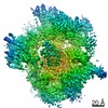 11694MC 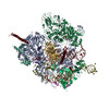 7aavC 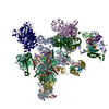 7abgC 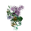 7abhC 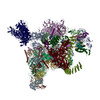 7abiC M: map data used to model this data C: citing same article ( |
|---|---|
| Similar structure data | |
| EM raw data | EMPIAR-10616 (Title: Cryo-EM dataset of human pre-Bact spliceosome / Data size: 584.5 Data #1: Motion-corrected micrographs (without dose-weighting) of human pre-Bact spliceosome [micrographs - single frame] Data #2: Motion-corrected micrographs (with dose-weighting) of human pre-Bact spliceosome [micrographs - single frame]) |


-Links
 PDBj
PDBj


- Assembly
| Deposited unit | 
|
|---|---|
| 1 |
|
-Components
-Protein , 12 types, 12 molecules QIArNqRXvGKA4
| #1: Protein | Mass: 17032.850 Da / Num. of mol.: 1 / Source method: isolated from a natural source / Source: (natural) Homo sapiens (human) / Cell line: Hela / References: UniProt: P41223 |
|---|---|
| #2: Protein | Mass: 37563.863 Da / Num. of mol.: 1 / Source method: isolated from a natural source / Source: (natural) Homo sapiens (human) / Cell line: Hela / References: UniProt: Q8NAV1 |
| #3: Protein | Mass: 273974.250 Da / Num. of mol.: 1 / Source method: isolated from a natural source / Source: (natural) Homo sapiens (human) / Cell line: Hela / References: UniProt: Q6P2Q9 |
| #4: Protein | Mass: 109560.625 Da / Num. of mol.: 1 / Source method: isolated from a natural source / Source: (natural) Homo sapiens (human) / Cell line: Hela / References: UniProt: Q15029 |
| #5: Protein | Mass: 23664.047 Da / Num. of mol.: 1 / Source method: isolated from a natural source / Source: (natural) Homo sapiens (human) / Cell line: Hela / References: UniProt: Q96NC0 |
| #6: Protein | Mass: 8560.945 Da / Num. of mol.: 1 / Source method: isolated from a natural source / Source: (natural) Homo sapiens (human) / Cell line: Hela / References: UniProt: Q9BZL1 |
| #7: Protein | Mass: 26674.447 Da / Num. of mol.: 1 / Source method: isolated from a natural source / Source: (natural) Homo sapiens (human) / Cell line: Hela / References: UniProt: Q9P013 |
| #10: Protein | Mass: 70098.641 Da / Num. of mol.: 1 / Source method: isolated from a natural source / Source: (natural) Homo sapiens (human) / Cell line: Hela / References: UniProt: Q9Y2W2 |
| #11: Protein | Mass: 61610.703 Da / Num. of mol.: 1 / Source method: isolated from a natural source / Source: (natural) Homo sapiens (human) / Cell line: Hela / References: UniProt: Q13573 |
| #12: Protein | Mass: 57280.758 Da / Num. of mol.: 1 / Source method: isolated from a natural source / Source: (natural) Homo sapiens (human) / Cell line: Hela / References: UniProt: O43660 |
| #14: Protein | Mass: 52050.527 Da / Num. of mol.: 1 / Source method: isolated from a natural source / Source: (natural) Homo sapiens (human) / Cell line: Hela / References: UniProt: P55081 |
| #15: Protein | Mass: 121870.320 Da / Num. of mol.: 1 / Source method: isolated from a natural source / Source: (natural) Homo sapiens (human) / Cell line: Hela / References: UniProt: O14776 |
-RNA chain , 3 types, 3 molecules 56Z
| #8: RNA chain | Mass: 36908.668 Da / Num. of mol.: 1 / Source method: isolated from a natural source / Source: (natural) Homo sapiens (human) / Cell line: Hela / References: GenBank: 36515 |
|---|---|
| #9: RNA chain | Mass: 34098.270 Da / Num. of mol.: 1 / Source method: isolated from a natural source / Source: (natural) Homo sapiens (human) / Cell line: Hela |
| #13: RNA chain | Mass: 73712.359 Da / Num. of mol.: 1 / Source method: obtained synthetically / Source: (synth.) synthetic construct (others) |
-Non-polymers , 3 types, 3 molecules 


| #16: Chemical | ChemComp-IHP /  Mass: 660.035 Da / Num. of mol.: 1 / Source method: obtained synthetically / Formula: C6H18O24P6 Mass: 660.035 Da / Num. of mol.: 1 / Source method: obtained synthetically / Formula: C6H18O24P6 |
|---|---|
| #17: Chemical | ChemComp-GTP /  Mass: 523.180 Da / Num. of mol.: 1 / Source method: obtained synthetically / Formula: C10H16N5O14P3 / Comment: GTP, energy-carrying molecule*YM Mass: 523.180 Da / Num. of mol.: 1 / Source method: obtained synthetically / Formula: C10H16N5O14P3 / Comment: GTP, energy-carrying molecule*YM |
| #18: Chemical | ChemComp-MG /  Mass: 24.305 Da / Num. of mol.: 1 / Source method: obtained synthetically / Formula: Mg Mass: 24.305 Da / Num. of mol.: 1 / Source method: obtained synthetically / Formula: Mg |
-Details
| Has ligand of interest | N |
|---|---|
| Has protein modification | N |
-Experimental details
-Experiment
| Experiment | Method: ELECTRON MICROSCOPY |
|---|---|
| EM experiment | Aggregation state: PARTICLE / 3D reconstruction method: single particle reconstruction |
- Sample preparation
| Component |
| ||||||||||||||||||||||||
|---|---|---|---|---|---|---|---|---|---|---|---|---|---|---|---|---|---|---|---|---|---|---|---|---|---|
| Molecular weight | Units: MEGADALTONS / Experimental value: NO | ||||||||||||||||||||||||
| Source (natural) |
| ||||||||||||||||||||||||
| Source (recombinant) | Organism: synthetic construct (others) | ||||||||||||||||||||||||
| Buffer solution | pH: 7.9 | ||||||||||||||||||||||||
| Specimen | Embedding applied: NO / Shadowing applied: NO / Staining applied: NO / Vitrification applied: YES | ||||||||||||||||||||||||
| Specimen support | Grid material: COPPER / Grid type: Quantifoil R3.5/1 | ||||||||||||||||||||||||
| Vitrification | Instrument: FEI VITROBOT MARK IV / Cryogen name: ETHANE |
- Electron microscopy imaging
| Experimental equipment |  Model: Titan Krios / Image courtesy: FEI Company |
|---|---|
| Microscopy | Model: FEI TITAN KRIOS |
| Electron gun | Electron source:  FIELD EMISSION GUN / Accelerating voltage: 300 kV / Illumination mode: SPOT SCAN FIELD EMISSION GUN / Accelerating voltage: 300 kV / Illumination mode: SPOT SCAN |
| Electron lens | Mode: BRIGHT FIELD |
| Image recording | Average exposure time: 1 sec. / Electron dose: 2.25 e/Å2 / Detector mode: INTEGRATING / Film or detector model: FEI FALCON III (4k x 4k) |
| Image scans | Width: 4096 / Height: 4096 |
- Processing
| EM software |
| ||||||||||||||||||||
|---|---|---|---|---|---|---|---|---|---|---|---|---|---|---|---|---|---|---|---|---|---|
| CTF correction | Type: PHASE FLIPPING AND AMPLITUDE CORRECTION | ||||||||||||||||||||
| Symmetry | Point symmetry: C1 (asymmetric) | ||||||||||||||||||||
| 3D reconstruction | Resolution: 3.9 Å / Resolution method: FSC 0.143 CUT-OFF / Num. of particles: 84539 / Symmetry type: POINT | ||||||||||||||||||||
| Atomic model building | Protocol: RIGID BODY FIT / Space: REAL |
