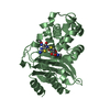
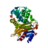
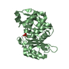
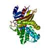
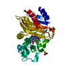
beta-lactam antibiotic catabolic process / beta-lactamase activity / beta-lactamase / periplasmic space / response to antibiotic / extracellular region Similarity search - Function
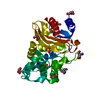
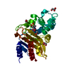
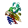
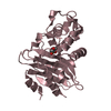
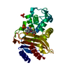
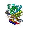
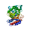
Beta-lactamase, class-A active site / Beta-lactamase class-A active site. / Beta-lactamase class A, catalytic domain / Beta-lactamase enzyme family / Beta-lactamase, class-A / Beta-lactamase/transpeptidase-like Similarity search - Domain/homology
Resolution: 1.4→44.84 Å / Cor.coef. Fo:Fc: 0.976 / Cor.coef. Fo:Fc free: 0.968 / SU B: 2.373 / SU ML: 0.04 / Cross valid method: THROUGHOUT / ESU R: 0.059 / ESU R Free: 0.056 / Stereochemistry target values: MAXIMUM LIKELIHOOD / Details: HYDROGENS HAVE BEEN ADDED IN THE RIDING POSITIONS
Rfactor
Num. reflection
% reflection
Selection details
Rfree
0.17229
2145
4.7 %
RANDOM
Rwork
0.13453
-
-
-
obs
0.13634
43191
97.44 %
-
Solvent computation
Ion probe radii: 0.8 Å / Shrinkage radii: 0.8 Å / VDW probe radii: 1.2 Å / Solvent model: MASK
 Movie
Movie Controller


 Open data
 Basic information
 Components
 Keywords
 Function and homology information
 Mycobacterium tuberculosis (bacteria)
 X-RAY DIFFRACTION /
  Authors
 Netherlands, 2items
  Citation
 Structure visualization
 Downloads & links
 Other downloads
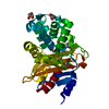


 PDBj
PDBj


 Assembly
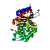

 Mass: 62.068 Da / Num. of mol.: 5 / Source method: obtained synthetically / Formula: C2H6O2
 Mass: 18.015 Da / Num. of mol.: 156 / Source method: isolated from a natural source / Formula: H2O
 Sample preparation
 / Beamline: X06SA / Wavelength: 1 Å
 Processing