 Movie
Movie Controller

[English] 日本語
 Yorodumi
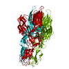
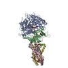
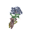
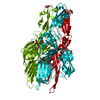
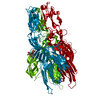
- PDB-7a4a: Envelope glycprotein of endogenous retrovirus Y032 (Atlas virus) ... -
+ Open data

- Basic information
| Entry | Database: PDB / ID: 7a4a | ||||||||||||
|---|---|---|---|---|---|---|---|---|---|---|---|---|---|
| Title | Envelope glycprotein of endogenous retrovirus Y032 (Atlas virus) from the human hookworm Ancylostoma ceylanicum | ||||||||||||
 Components Components | Integrase catalytic domain-containing protein | ||||||||||||
 Keywords Keywords | VIRAL PROTEIN / class II membrane fusion protein / retroviral envelope protein (Env) / lipid binding protein / disulfide bonding | ||||||||||||
| Function / homology |  Function and homology information Function and homology informationDNA polymerase complex / DNA integration / nucleic acid binding / DNA repair Similarity search - Function | ||||||||||||
| Biological species |  Ancylostoma ceylanicum (invertebrata) Ancylostoma ceylanicum (invertebrata) | ||||||||||||
| Method | ELECTRON MICROSCOPY / single particle reconstruction / cryo EM / Resolution: 3.76 Å | ||||||||||||
 Authors Authors | Mata, C.P. / Merchant, M. / Modis, Y. | ||||||||||||
| Funding support |  United Kingdom, United Kingdom,  United States, 3items United States, 3items
| ||||||||||||
 Citation Citation | Journal: Sci Adv / Year: 2022 Title: A bioactive phlebovirus-like envelope protein in a hookworm endogenous virus. Authors: Monique Merchant / Carlos P Mata / Yangci Liu / Haoming Zhai / Anna V Protasio / Yorgo Modis / Abstract: Endogenous viral elements (EVEs), accounting for 15% of our genome, serve as a genetic reservoir from which new genes can emerge. Nematode EVEs are particularly diverse and informative of virus ...Endogenous viral elements (EVEs), accounting for 15% of our genome, serve as a genetic reservoir from which new genes can emerge. Nematode EVEs are particularly diverse and informative of virus evolution. We identify Atlas virus-an intact retrovirus-like EVE in the human hookworm , with an envelope protein genetically related to G-G glycoproteins from the family Phenuiviridae. A cryo-EM structure of Atlas G reveals a class II viral membrane fusion protein fold not previously seen in retroviruses. Atlas G has the structural hallmarks of an active fusogen. Atlas G trimers insert into membranes with endosomal lipid compositions and low pH. When expressed on the plasma membrane, Atlas G has cell-cell fusion activity. With its preserved biological activities, Atlas G has the potential to acquire a cellular function. Our work reveals structural plasticity in reverse-transcribing RNA viruses. #1: Journal: Biorxiv / Year: 2021Title: A bioactive phlebovirus-like envelope protein in a hookworm endogenous virus Authors: Merchant, M. / Mata, C.P. / Liu, Y. / Zhai, H. / Protasio, A.V. / Modis, Y. | ||||||||||||
| History |
|
- Structure visualization
| Movie |
Movie viewer |
|---|---|
| Structure viewer | Molecule: MolmilJmol/JSmol |
- Downloads & links
-Download
| PDBx/mmCIF format | 7a4a.cif.gz | 419.8 KB | Display | PDBx/mmCIF format |
|---|---|---|---|---|
| PDB format | pdb7a4a.ent.gz | 346.8 KB | Display | PDB format |
| PDBx/mmJSON format | 7a4a.json.gz | Tree view | PDBx/mmJSON format | |
| Others |  Other downloads Other downloads |
-Validation report
| Arichive directory | https://data.pdbj.org/pub/pdb/validation_reports/a4/7a4aftp://data.pdbj.org/pub/pdb/validation_reports/a4/7a4a | HTTPS FTP |
|---|
-Related structure data
| Related structure data |  11630MC M: map data used to model this data C: citing same article ( |
|---|---|
| Similar structure data | |
| EM raw data | EMPIAR-10266 (Title: CryoEM image reconstuction of the envelope protein of endogenous retrovirus Y032 from the human hookworm Ancylostoma ceylanicum Data size: 5.6 TB Data #1: Unaligned multi-frame micrographs of Env protein endogenous retrovirus AceY032 from Ancylostoma ceylanicum [micrographs - multiframe]) |
| Experimental dataset #1 | Data reference: 10.6019/EMPIAR-10266 / Data set type: EMPIAR |


-Links
 PDBj
PDBj
- Assembly
| Deposited unit | 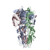
|
|---|---|
| 1 |
|
-Components
| #1: Protein | Mass: 51519.090 Da / Num. of mol.: 3 Source method: isolated from a genetically manipulated source Details: In chains A, B and C, residue Asn414 is covalently modified with an N-linked N-acetyl glucosamine ligand. Chains A, B and C each contain the following 15 disulfide bonds: Cys1-Cys41, Cys14- ...Details: In chains A, B and C, residue Asn414 is covalently modified with an N-linked N-acetyl glucosamine ligand. Chains A, B and C each contain the following 15 disulfide bonds: Cys1-Cys41, Cys14-Cys23, Cys66-Cys162, Cys87-Cys135, Cys93-Cys142, Cys98-Cys123, Cys127-Cys132, Cys129-Cys138, Cys246-Cys257, Cys264-Cys277, Cys266-Cys275, Cys337-Cys408, Cys347-Cys350, Cys360-Cys382, Cys373-Cys404. Source: (gene. exp.) Ancylostoma ceylanicum (invertebrata) / Gene: Acey_s0020.g108, Y032_0020g108 / Plasmid: pMT/BiP/V5-His / Cell line (production host): d.mel-2 / Production host:  #2: Sugar |   Type: D-saccharide, beta linking / Mass: 221.208 Da / Num. of mol.: 3 Type: D-saccharide, beta linking / Mass: 221.208 Da / Num. of mol.: 3Source method: isolated from a genetically manipulated source Formula: C8H15NO6 Has ligand of interest | N | Has protein modification | Y | |
|---|
-Experimental details
-Experiment
| Experiment | Method: ELECTRON MICROSCOPY |
|---|---|
| EM experiment | Aggregation state: PARTICLE / 3D reconstruction method: single particle reconstruction |
- Sample preparation
| Component | Name: Viral envelope glycoprotein / Type: COMPLEX Details: Envelope glycoprotein of endogenous retrovirus Y032 (Atlas virus) from Ancylostoma ceylanicum Entity ID: #1 / Source: RECOMBINANT | |||||||||||||||||||||||||
|---|---|---|---|---|---|---|---|---|---|---|---|---|---|---|---|---|---|---|---|---|---|---|---|---|---|---|
| Molecular weight | Value: 0.143 MDa / Experimental value: YES | |||||||||||||||||||||||||
| Source (natural) | Organism: Ancylostoma ceylanicum (invertebrata) | |||||||||||||||||||||||||
| Source (recombinant) | Organism: | |||||||||||||||||||||||||
| Buffer solution | pH: 8 Details: 20 mM Tris/HCl (NH12C4O3Cl) 0.1 M NaCl 'sodium chloride' 5 % glycerol (C3H8O3) 0.5 mM TCEP (C9H15O6P) | |||||||||||||||||||||||||
| Buffer component |
| |||||||||||||||||||||||||
| Specimen | Conc.: 0.025 mg/ml / Embedding applied: NO / Shadowing applied: NO / Staining applied: NO / Vitrification applied: YES | |||||||||||||||||||||||||
| Specimen support | Grid material: COPPER / Grid mesh size: 400 divisions/in. / Grid type: Quantifoil R1.2/1.3 | |||||||||||||||||||||||||
| Vitrification | Instrument: FEI VITROBOT MARK IV / Cryogen name: ETHANE / Humidity: 100 % / Chamber temperature: 277.2 K / Details: Grids were blotted for 4 s |
- Electron microscopy imaging
| Experimental equipment |  Model: Titan Krios / Image courtesy: FEI Company |
|---|---|
| Microscopy | Model: FEI TITAN KRIOS |
| Electron gun | Electron source:  FIELD EMISSION GUN / Accelerating voltage: 300 kV / Illumination mode: FLOOD BEAM FIELD EMISSION GUN / Accelerating voltage: 300 kV / Illumination mode: FLOOD BEAM |
| Electron lens | Mode: BRIGHT FIELD / Calibrated magnification: 75000 X / Nominal defocus max: 3500 nm / Nominal defocus min: 1300 nm / Alignment procedure: ZEMLIN TABLEAU |
| Specimen holder | Cryogen: NITROGEN / Specimen holder model: FEI TITAN KRIOS AUTOGRID HOLDER |
| Image recording | Average exposure time: 8 sec. / Electron dose: 46.18 e/Å2 / Detector mode: COUNTING / Film or detector model: GATAN K2 SUMMIT (4k x 4k) / Num. of grids imaged: 1 / Num. of real images: 3027 / Details: Dose rate = 1.28 e- A^-2 per frame |
| EM imaging optics | Energyfilter name: GIF Quantum LS / Chromatic aberration corrector: None / Energyfilter slit width: 20 eV / Phase plate: OTHER / Spherical aberration corrector: None |
| Image scans | Sampling size: 5 µm / Width: 3838 / Height: 3710 / Movie frames/image: 36 |
- Processing
| Software | Name: PHENIX / Version: 1.17.1_3660: / Classification: refinement | ||||||||||||||||||||||||||||||||||||||||
|---|---|---|---|---|---|---|---|---|---|---|---|---|---|---|---|---|---|---|---|---|---|---|---|---|---|---|---|---|---|---|---|---|---|---|---|---|---|---|---|---|---|
| EM software |
| ||||||||||||||||||||||||||||||||||||||||
| Image processing | Details: Movies were motion-corrected and dose-weighted with MOTIONCOR2. | ||||||||||||||||||||||||||||||||||||||||
| CTF correction | Details: Aligned, non-dose-weighted micrographs were then used to estimate the CTF. Type: PHASE FLIPPING AND AMPLITUDE CORRECTION | ||||||||||||||||||||||||||||||||||||||||
| Particle selection | Num. of particles selected: 987570 Details: 2D references from initial datasets were used to auto-pick the micrographs. One round of reference-free 2D classification was performed to produce templates for better reference-dependent auto-picking. | ||||||||||||||||||||||||||||||||||||||||
| Symmetry | Point symmetry: C3 (3 fold cyclic) | ||||||||||||||||||||||||||||||||||||||||
| 3D reconstruction | Resolution: 3.76 Å / Resolution method: FSC 0.143 CUT-OFF / Num. of particles: 197145 / Algorithm: FOURIER SPACE / Num. of class averages: 1 / Symmetry type: POINT | ||||||||||||||||||||||||||||||||||||||||
| Atomic model building | B value: 82 / Protocol: FLEXIBLE FIT / Space: REAL / Target criteria: Cross-correlation coefficient Details: A homology model was built from PDB:6EGU using the Swiss-Model server (swissmodel.expasy.org). The model was docked as a rigid body into the density with UCSF Chimera prior to refinement. | ||||||||||||||||||||||||||||||||||||||||
| Atomic model building | PDB-ID: 6EGU Pdb chain-ID: A / Accession code: 6EGU Details: A homology model was built from PDB:6EGU using the Swiss-Model server (swissmodel.expasy.org). The model was docked as a rigid body into the density with UCSF Chimera prior to refinement. Pdb chain residue range: 691-1136 / Source name: PDB / Type: experimental model | ||||||||||||||||||||||||||||||||||||||||
| Refine LS restraints |
|

