 Movie
Movie Controller

[English] 日本語
 Yorodumi
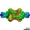
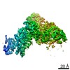
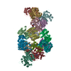
- PDB-7a1d: Cryo-EM map of the large glutamate dehydrogenase composed of 180 ... -
+ Open data

- Basic information
| Entry | Database: PDB / ID: 7a1d | |||||||||||||||||||||||||||||||||||||||||||||||||||||||||||||||||||||||||||||||||
|---|---|---|---|---|---|---|---|---|---|---|---|---|---|---|---|---|---|---|---|---|---|---|---|---|---|---|---|---|---|---|---|---|---|---|---|---|---|---|---|---|---|---|---|---|---|---|---|---|---|---|---|---|---|---|---|---|---|---|---|---|---|---|---|---|---|---|---|---|---|---|---|---|---|---|---|---|---|---|---|---|---|---|
| Title | Cryo-EM map of the large glutamate dehydrogenase composed of 180 kDa subunits from Mycobacterium smegmatis (open conformation) | |||||||||||||||||||||||||||||||||||||||||||||||||||||||||||||||||||||||||||||||||
 Components Components | NAD-specific glutamate dehydrogenase | |||||||||||||||||||||||||||||||||||||||||||||||||||||||||||||||||||||||||||||||||
 Keywords Keywords | OXIDOREDUCTASE / large glutamate dehydrogenase Mycobacterium metabolism | |||||||||||||||||||||||||||||||||||||||||||||||||||||||||||||||||||||||||||||||||
| Function / homology |  Function and homology information Function and homology informationglutamate dehydrogenase / L-glutamate dehydrogenase (NAD+) activity / L-glutamate catabolic process / L-aspartate:2-oxoglutarate transaminase activity Similarity search - Function | |||||||||||||||||||||||||||||||||||||||||||||||||||||||||||||||||||||||||||||||||
| Biological species |  Mycolicibacterium smegmatis MC2 155 (bacteria) Mycolicibacterium smegmatis MC2 155 (bacteria) | |||||||||||||||||||||||||||||||||||||||||||||||||||||||||||||||||||||||||||||||||
| Method | ELECTRON MICROSCOPY / single particle reconstruction / cryo EM / Resolution: 4.19 Å | |||||||||||||||||||||||||||||||||||||||||||||||||||||||||||||||||||||||||||||||||
 Authors Authors | Lazaro, M. / Melero, R. / Huet, C. / Lopez-Alonso, J.P. / Delgado, S. / Dodu, A. / Bruch, E.M. / Abriata, L.A. / Alzari, P.M. / Valle, M. / Lisa, M.N. | |||||||||||||||||||||||||||||||||||||||||||||||||||||||||||||||||||||||||||||||||
| Funding support |  Spain, 1items Spain, 1items
| |||||||||||||||||||||||||||||||||||||||||||||||||||||||||||||||||||||||||||||||||
 Citation Citation | Journal: Commun Biol / Year: 2021 Title: 3D architecture and structural flexibility revealed in the subfamily of large glutamate dehydrogenases by a mycobacterial enzyme. Authors: Melisa Lázaro / Roberto Melero / Charlotte Huet / Jorge P López-Alonso / Sandra Delgado / Alexandra Dodu / Eduardo M Bruch / Luciano A Abriata / Pedro M Alzari / Mikel Valle / María-Natalia Lisa /    Abstract: Glutamate dehydrogenases (GDHs) are widespread metabolic enzymes that play key roles in nitrogen homeostasis. Large glutamate dehydrogenases composed of 180 kDa subunits (L-GDHs) contain long N- ...Glutamate dehydrogenases (GDHs) are widespread metabolic enzymes that play key roles in nitrogen homeostasis. Large glutamate dehydrogenases composed of 180 kDa subunits (L-GDHs) contain long N- and C-terminal segments flanking the catalytic core. Despite the relevance of L-GDHs in bacterial physiology, the lack of structural data for these enzymes has limited the progress of functional studies. Here we show that the mycobacterial L-GDH (mL-GDH) adopts a quaternary structure that is radically different from that of related low molecular weight enzymes. Intersubunit contacts in mL-GDH involve a C-terminal domain that we propose as a new fold and a flexible N-terminal segment comprising ACT-like and PAS-type domains that could act as metabolic sensors for allosteric regulation. These findings uncover unique aspects of the structure-function relationship in the subfamily of L-GDHs. | |||||||||||||||||||||||||||||||||||||||||||||||||||||||||||||||||||||||||||||||||
| History |
|
- Structure visualization
| Movie |
Movie viewer |
|---|---|
| Structure viewer | Molecule: MolmilJmol/JSmol |
- Downloads & links
-Download
| PDBx/mmCIF format | 7a1d.cif.gz | 792.8 KB | Display | PDBx/mmCIF format |
|---|---|---|---|---|
| PDB format | pdb7a1d.ent.gz | 609.7 KB | Display | PDB format |
| PDBx/mmJSON format | 7a1d.json.gz | Tree view | PDBx/mmJSON format | |
| Others |  Other downloads Other downloads |
-Validation report
| Arichive directory | https://data.pdbj.org/pub/pdb/validation_reports/a1/7a1dftp://data.pdbj.org/pub/pdb/validation_reports/a1/7a1d | HTTPS FTP |
|---|
-Related structure data
| Related structure data | 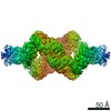 11606MC  7jsrC M: map data used to model this data C: citing same article ( |
|---|---|
| Similar structure data |


-Links
 PDBj
PDBj- Assembly
| Deposited unit | 
|
|---|---|
| 1 |
|
-Components
| #1: Protein | Mass: 176341.859 Da / Num. of mol.: 4 Source method: isolated from a genetically manipulated source Source: (gene. exp.) Mycolicibacterium smegmatis MC2 155 (bacteria)Strain: ATCC 700084 / mc(2)155 / Gene: gdh, MSMEG_4699, MSMEI_4582 / Production host: Has protein modification | N | |
|---|
-Experimental details
-Experiment
| Experiment | Method: ELECTRON MICROSCOPY |
|---|---|
| EM experiment | Aggregation state: PARTICLE / 3D reconstruction method: single particle reconstruction |
- Sample preparation
| Component | Name: Cryo-EM map of the large glutamate dehydrogenase composed of 180 kDa subunits from Mycobacterium smegmatis (open conformation) Type: COMPLEX / Entity ID: all / Source: RECOMBINANT |
|---|---|
| Molecular weight | Value: 0.705 MDa / Experimental value: NO |
| Source (natural) | Organism: Mycolicibacterium smegmatis MC2 155 (bacteria) |
| Source (recombinant) | Organism: |
| Buffer solution | pH: 6 |
| Specimen | Conc.: 0.3 mg/ml / Embedding applied: NO / Shadowing applied: NO / Staining applied: NO / Vitrification applied: YES |
| Specimen support | Grid material: COPPER / Grid mesh size: 300 divisions/in. / Grid type: Quantifoil |
| Vitrification | Cryogen name: ETHANE |
- Electron microscopy imaging
| Experimental equipment |  Model: Titan Krios / Image courtesy: FEI Company |
|---|---|
| Microscopy | Model: TFS KRIOS |
| Electron gun | Electron source:  FIELD EMISSION GUN / Accelerating voltage: 300 kV / Illumination mode: FLOOD BEAM FIELD EMISSION GUN / Accelerating voltage: 300 kV / Illumination mode: FLOOD BEAM |
| Electron lens | Mode: BRIGHT FIELD / Calibrated magnification: 47170 X / Nominal defocus max: 3260 nm / Nominal defocus min: 670 nm / Calibrated defocus min: 670 nm / Calibrated defocus max: 3260 nm / Cs: 2.7 mm |
| Image recording | Electron dose: 40 e/Å2 / Detector mode: COUNTING / Film or detector model: GATAN K2 SUMMIT (4k x 4k) |
| Image scans | Movie frames/image: 20 / Used frames/image: 1-20 |
- Processing
| EM software | Name: PHENIX / Version: 1.21.1_5286 / Category: model refinement |
|---|---|
| CTF correction | Type: PHASE FLIPPING ONLY |
| Symmetry | Point symmetry: D2 (2x2 fold dihedral) |
| 3D reconstruction | Resolution: 4.19 Å / Resolution method: FSC 0.143 CUT-OFF / Num. of particles: 63715 / Algorithm: FOURIER SPACE / Symmetry type: POINT |
| Atomic model building | Protocol: OTHER Details: Model fitting into the cryo-EM map was performed using the programs UCSF Chimera (Pettersen et al., 2004), Namdinator (Kidmose et al., 2019), phenix.real_space_refine (Afonine et al., 2018) ...Details: Model fitting into the cryo-EM map was performed using the programs UCSF Chimera (Pettersen et al., 2004), Namdinator (Kidmose et al., 2019), phenix.real_space_refine (Afonine et al., 2018) and Coot (Emsley, Lohkamp, Scott, & Cowtan, 2010). Residues 500-1588 from the crystal structure of Se-Met mL-GDH180 (PDB code 7JSR) were fitted into the cryo-EM map. Se-methionine residues were replaced by methionine residues using Coot (Emsley et al., 2010) and the model was finally refined employing phenix.real_space_refine (Afonine et al., 2018) with NCS and secondary structure restraints. |
| Refinement | Cross valid method: NONE |
