Type: MAR scanner 345 mm plate / Detector: IMAGE PLATE / Date: Sep 30, 2010
Radiation
Protocol: SINGLE WAVELENGTH / Monochromatic (M) / Laue (L): M / Scattering type: x-ray
Radiation wavelength
Wavelength: 1.54187 Å / Relative weight: 1
Reflection
Resolution: 1.939→43.187 Å / Num. obs: 23907 / % possible obs: 91 % / Redundancy: 3.4 % / Rpim(I) all: 0.071 / Rrim(I) all: 0.133 / Rsym value: 0.112 / Net I/av σ(I): 5.8 / Net I/σ(I): 8.1
Reflection shell
Diffraction-ID: 1
Resolution (Å)
Redundancy (%)
Rmerge(I) obs
Mean I/σ(I) obs
Num. unique obs
Rpim(I) all
Rrim(I) all
Rsym value
% possible all
1.939-2.03
3.3
0.58
1.3
2905
0.372
0.69
0.58
76.2
2.03-2.15
3.4
0.364
2
3292
0.232
0.432
0.364
91.2
2.15-2.3
3.4
0.267
2.7
3131
0.17
0.317
0.267
92.1
2.3-2.49
3.4
0.215
3.4
2944
0.137
0.255
0.215
92.6
2.49-2.73
3.4
0.163
4.4
2709
0.104
0.194
0.163
93.4
2.73-3.05
3.4
0.113
6.3
2489
0.072
0.135
0.113
94.2
3.05-3.52
3.4
0.075
9.2
2220
0.048
0.089
0.075
95
3.52-4.31
3.4
0.059
10.6
1893
0.038
0.07
0.059
95.9
4.31-6.09
3.3
0.057
10.3
1488
0.036
0.067
0.057
96.2
6.09-43.187
3.2
0.053
10.9
836
0.034
0.063
0.053
97
-
Phasing
Phasing
Method: molecular replacement
-
Processing
Software
Name
Version
Classification
NB
MOSFLM
datareduction
SCALA
3.3.15
datascaling
PHASER
phasing
REFMAC
5.8.0158
refinement
PDB_EXTRACT
3.25
dataextraction
Refinement
Method to determine structure: MOLECULAR REPLACEMENT Starting model: inhouse Resolution: 1.94→43.187 Å / Cor.coef. Fo:Fc: 0.959 / Cor.coef. Fo:Fc free: 0.921 / SU B: 7.284 / SU ML: 0.107 / SU R Cruickshank DPI: 0.1553 / Cross valid method: THROUGHOUT / σ(F): 0 / ESU R: 0.155 / ESU R Free: 0.147 / Stereochemistry target values: MAXIMUM LIKELIHOOD Details: HYDROGENS HAVE BEEN ADDED IN THE RIDING POSITIONS U VALUES : WITH TLS ADDED
Rfactor
Num. reflection
% reflection
Selection details
Rfree
0.212
1221
5.1 %
RANDOM
Rwork
0.1637
-
-
-
obs
0.1662
22685
92.37 %
-
Solvent computation
Ion probe radii: 0.8 Å / Shrinkage radii: 0.8 Å / VDW probe radii: 1.2 Å / Solvent model: BABINET MODEL WITH MASK
In the structure databanks used in Yorodumi, some data are registered as the other names, "COVID-19 virus" and "2019-nCoV". Here are the details of the virus and the list of structure data.
Jan 31, 2019. EMDB accession codes are about to change! (news from PDBe EMDB page)
EMDB accession codes are about to change! (news from PDBe EMDB page)
The allocation of 4 digits for EMDB accession codes will soon come to an end. Whilst these codes will remain in use, new EMDB accession codes will include an additional digit and will expand incrementally as the available range of codes is exhausted. The current 4-digit format prefixed with “EMD-” (i.e. EMD-XXXX) will advance to a 5-digit format (i.e. EMD-XXXXX), and so on. It is currently estimated that the 4-digit codes will be depleted around Spring 2019, at which point the 5-digit format will come into force.
The EM Navigator/Yorodumi systems omit the EMD- prefix.
Related info.:Q: What is EMD? / ID/Accession-code notation in Yorodumi/EM Navigator
Yorodumi is a browser for structure data from EMDB, PDB, SASBDB, etc.
This page is also the successor to EM Navigator detail page, and also detail information page/front-end page for Omokage search.
The word "yorodu" (or yorozu) is an old Japanese word meaning "ten thousand". "mi" (miru) is to see.
Related info.:EMDB / PDB / SASBDB / Comparison of 3 databanks / Yorodumi Search / Aug 31, 2016. New EM Navigator & Yorodumi / Yorodumi Papers / Jmol/JSmol / Function and homology information / Changes in new EM Navigator and Yorodumi
 Movie
Movie Controller


 Open data
 Basic information
 Components
 Keywords
 Function and homology information
 Homo sapiens (human)
 Hirudo medicinalis (medicinal leech)
 X-RAY DIFFRACTION /
  Authors
 Citation
 Structure visualization
 Downloads & links
 Other downloads


 PDBj
PDBj


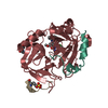
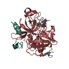
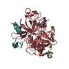
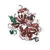
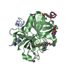
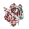
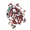
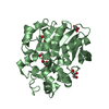
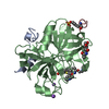
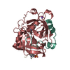
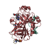
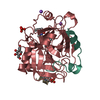
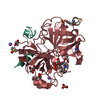
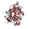
 Assembly
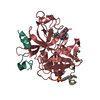

 Type: D-saccharide, beta linking / Mass: 221.208 Da / Num. of mol.: 1 / Source method: obtained synthetically / Formula: C8H15NO6
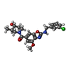

 Mass: 429.897 Da / Num. of mol.: 1 / Source method: obtained synthetically / Formula: C22H24ClN3O4 / Feature type: SUBJECT OF INVESTIGATION
 Sample preparation
 Processing