 Movie
Movie Controller


+ Open data

- Basic information
| Entry | Database: PDB / ID: 6zs1 | ||||||
|---|---|---|---|---|---|---|---|
| Title | Chaetomium thermophilum CuZn-superoxide dismutase | ||||||
 Components Components | Superoxide dismutase [Cu-Zn] | ||||||
 Keywords Keywords | METAL BINDING PROTEIN / thermophilic fungus / superoxide / antioxidant / thermostability / metalloenzyme / metal-binding protein | ||||||
| Function / homology |  Function and homology information Function and homology informationsuperoxide dismutase / superoxide dismutase activity / copper ion binding Similarity search - Function | ||||||
| Biological species |  Chaetomium thermophilum var. thermophilum DSM 1495 (fungus) Chaetomium thermophilum var. thermophilum DSM 1495 (fungus) | ||||||
| Method |  X-RAY DIFFRACTION / SYNCHROTRON / MOLECULAR REPLACEMENT / Resolution: 1.56 Å X-RAY DIFFRACTION / SYNCHROTRON / MOLECULAR REPLACEMENT / Resolution: 1.56 Å | ||||||
 Authors Authors | Papageorgiou, A.C. / Mohsin, I. | ||||||
 Citation Citation | Journal: Protein Pept.Lett. / Year: 2021 Title: Crystal Structure of a Cu,Zn Superoxide Dismutase From the Thermophilic Fungus Chaetomium thermophilum. Authors: Mohsin, I. / Zhang, L.Q. / Li, D.C. / Papageorgiou, A.C. #1: Journal: Acta Crystallogr. Sect. F Struct. Biol. Cryst. Commun. Year: 2010 Title: Expression, purification and crystallization of Chaetomium thermophilum Cu,Zn superoxide dismutase. Authors: Wakadkar, S. / Zhang, L.-Q. / Li, D.-C. / Haikarainen, T. / Dhavala, P. / Papageorgiou, A.C. | ||||||
| History |
|
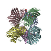
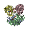
- Structure visualization
| Structure viewer | Molecule: MolmilJmol/JSmol |
|---|
- Downloads & links
-Download
| PDBx/mmCIF format | 6zs1.cif.gz | 369.8 KB | Display | PDBx/mmCIF format |
|---|---|---|---|---|
| PDB format | pdb6zs1.ent.gz | 239.8 KB | Display | PDB format |
| PDBx/mmJSON format | 6zs1.json.gz | Tree view | PDBx/mmJSON format | |
| Others |  Other downloads Other downloads |
-Validation report
| Arichive directory | https://data.pdbj.org/pub/pdb/validation_reports/zs/6zs1ftp://data.pdbj.org/pub/pdb/validation_reports/zs/6zs1 | HTTPS FTP |
|---|
-Related structure data
| Related structure data |  1f1gS S: Starting model for refinement |
|---|---|
| Similar structure data |


-Links
 PDBj
PDBj
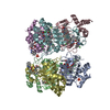
- Assembly
| Deposited unit | 
| ||||||||||||
|---|---|---|---|---|---|---|---|---|---|---|---|---|---|
| 1 |
| ||||||||||||
| Unit cell |
|
-Components
-Protein , 1 types, 8 molecules ABCDEFGH
| #1: Protein | Mass: 16369.033 Da / Num. of mol.: 8 Source method: isolated from a genetically manipulated source Details: There is clear density for Ile at residue 48 (coordinate file numbering) despite the original sequence contains a Thr at the same position YVE sequence at the N-terminal belongs to the ...Details: There is clear density for Ile at residue 48 (coordinate file numbering) despite the original sequence contains a Thr at the same position YVE sequence at the N-terminal belongs to the expression vector secretion signal Source: (gene. exp.) Chaetomium thermophilum var. thermophilum DSM 1495 (fungus)Gene: CTHT_0013440 / Production host: Komagataella phaffii GS115 (fungus) / References: UniProt: G0S1F8, superoxide dismutase |
|---|
-Non-polymers , 6 types, 2493 molecules 


| #2: Chemical | ChemComp-ZN /  Mass: 65.409 Da / Num. of mol.: 8 / Source method: isolated from a natural source / Formula: Zn / Feature type: SUBJECT OF INVESTIGATION Mass: 65.409 Da / Num. of mol.: 8 / Source method: isolated from a natural source / Formula: Zn / Feature type: SUBJECT OF INVESTIGATION#3: Chemical | ChemComp-PO4 /  Mass: 94.971 Da / Num. of mol.: 7 / Source method: obtained synthetically / Formula: PO4 Mass: 94.971 Da / Num. of mol.: 7 / Source method: obtained synthetically / Formula: PO4#4: Chemical | ChemComp-NA /  Mass: 22.990 Da / Num. of mol.: 15 / Source method: obtained synthetically / Formula: Na Mass: 22.990 Da / Num. of mol.: 15 / Source method: obtained synthetically / Formula: Na#5: Chemical | ChemComp-CU /  Mass: 63.546 Da / Num. of mol.: 4 / Source method: obtained synthetically / Formula: Cu / Feature type: SUBJECT OF INVESTIGATION Mass: 63.546 Da / Num. of mol.: 4 / Source method: obtained synthetically / Formula: Cu / Feature type: SUBJECT OF INVESTIGATION#6: Chemical | ChemComp-GOL /  Mass: 92.094 Da / Num. of mol.: 4 / Source method: obtained synthetically / Formula: C3H8O3 Mass: 92.094 Da / Num. of mol.: 4 / Source method: obtained synthetically / Formula: C3H8O3#7: Water | ChemComp-HOH / | Mass: 18.015 Da / Num. of mol.: 2455 / Source method: isolated from a natural source / Formula: H2O |
|---|
-Details
| Has ligand of interest | Y |
|---|---|
| Has protein modification | Y |
-Experimental details
-Experiment
| Experiment | Method: X-RAY DIFFRACTION / Number of used crystals: 1 |
|---|
- Sample preparation
| Crystal | Density Matthews: 2.76 Å3/Da / Density % sol: 55.9 % / Description: Bipyramids |
|---|---|
| Crystal grow | Temperature: 289 K / Method: vapor diffusion, hanging drop / pH: 8.2 / Details: NaK phosphate 1.2-1.4 M |
-Data collection
| Diffraction | Mean temperature: 100 K / Serial crystal experiment: N |
|---|---|
| Diffraction source | Source: SYNCHROTRON / Site: EMBL/DESY, HAMBURG  / Beamline: X11 / Wavelength: 0.8047 Å / Beamline: X11 / Wavelength: 0.8047 Å |
| Detector | Type: MAR555 FLAT PANEL / Detector: IMAGE PLATE / Date: Apr 10, 2010 |
| Radiation | Protocol: SINGLE WAVELENGTH / Monochromatic (M) / Laue (L): M / Scattering type: x-ray |
| Radiation wavelength | Wavelength: 0.8047 Å / Relative weight: 1 |
| Reflection | Resolution: 1.56→38.59 Å / Num. obs: 194568 / % possible obs: 98 % / Redundancy: 5 % / Biso Wilson estimate: 19.25 Å2 / CC1/2: 0.999 / Rrim(I) all: 0.008 / Net I/σ(I): 15 |
| Reflection shell | Resolution: 1.56→1.64 Å / Redundancy: 2.8 % / Mean I/σ(I) obs: 1.15 / Num. unique obs: 29651 / CC1/2: 0.416 / Rrim(I) all: 0.146 / % possible all: 92.2 |
- Processing
| Software |
| |||||||||||||||||||||||||||||||||||||||||||||||||||||||||||||||||||||||||||||
|---|---|---|---|---|---|---|---|---|---|---|---|---|---|---|---|---|---|---|---|---|---|---|---|---|---|---|---|---|---|---|---|---|---|---|---|---|---|---|---|---|---|---|---|---|---|---|---|---|---|---|---|---|---|---|---|---|---|---|---|---|---|---|---|---|---|---|---|---|---|---|---|---|---|---|---|---|---|---|
| Refinement | Method to determine structure: MOLECULAR REPLACEMENT Starting model: 1f1g Resolution: 1.56→38.59 Å / SU ML: 0.2459 / Cross valid method: FREE R-VALUE / σ(F): 1.35 / Phase error: 25.0058 Stereochemistry target values: GeoStd + Monomer Library + CDL v1.2
| |||||||||||||||||||||||||||||||||||||||||||||||||||||||||||||||||||||||||||||
| Solvent computation | Shrinkage radii: 0.9 Å / VDW probe radii: 1.11 Å / Solvent model: FLAT BULK SOLVENT MODEL | |||||||||||||||||||||||||||||||||||||||||||||||||||||||||||||||||||||||||||||
| Displacement parameters | Biso mean: 23.81 Å2 | |||||||||||||||||||||||||||||||||||||||||||||||||||||||||||||||||||||||||||||
| Refinement step | Cycle: LAST / Resolution: 1.56→38.59 Å
| |||||||||||||||||||||||||||||||||||||||||||||||||||||||||||||||||||||||||||||
| Refine LS restraints |
| |||||||||||||||||||||||||||||||||||||||||||||||||||||||||||||||||||||||||||||
| LS refinement shell |
|
