 Movie
Movie Controller


+ Open data

- Basic information
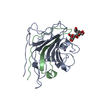
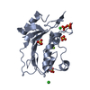
| Entry | Database: PDB / ID: 6zrw | ||||||
|---|---|---|---|---|---|---|---|
| Title | Crystal structure of the fungal lectin CML1 | ||||||
 Components Components | Mucin-binding lectin 1 | ||||||
 Keywords Keywords | CARBOHYDRATE / Lectin / fungi / Coprinopsis cinerea | ||||||
| Function / homology | carbohydrate binding / ACETATE ION / : / TRICHLOROPLATINATE / TETRACHLOROPLATINATE(II) / Mucin-binding lectin 1 Function and homology information Function and homology information | ||||||
| Biological species |  Coprinopsis cinerea (fungus) Coprinopsis cinerea (fungus) | ||||||
| Method |  X-RAY DIFFRACTION / SYNCHROTRON / SAD / Resolution: 1.35 Å X-RAY DIFFRACTION / SYNCHROTRON / SAD / Resolution: 1.35 Å | ||||||
 Authors Authors | Bleuler-Martinez, S. / Olieric, V. / Sharpe, M. / Capitani, G. / Aebi, M. / Kuenzler, M. | ||||||
| Funding support |  Switzerland, 1items Switzerland, 1items
| ||||||
 Citation Citation | Journal: Glycobiology / Year: 2022 Title: Structure-function relationship of a novel fucoside-binding fruiting body lectin from Coprinopsis cinerea exhibiting nematotoxic activity. Authors: Bleuler-Martinez, S. / Varrot, A. / Olieric, V. / Schubert, M. / Vogt, E. / Fetz, C. / Wohlschlager, T. / Plaza, D.F. / Walti, M. / Duport, Y. / Capitani, G. / Aebi, M. / Kunzler, M. | ||||||
| History |
|
- Structure visualization
| Structure viewer | Molecule: MolmilJmol/JSmol |
|---|
- Downloads & links
-Download
| PDBx/mmCIF format | 6zrw.cif.gz | 184.6 KB | Display | PDBx/mmCIF format |
|---|---|---|---|---|
| PDB format | pdb6zrw.ent.gz | 146.6 KB | Display | PDB format |
| PDBx/mmJSON format | 6zrw.json.gz | Tree view | PDBx/mmJSON format | |
| Others |  Other downloads Other downloads |
-Validation report
| Arichive directory | https://data.pdbj.org/pub/pdb/validation_reports/zr/6zrwftp://data.pdbj.org/pub/pdb/validation_reports/zr/6zrw | HTTPS FTP |
|---|
-Related structure data
| Related structure data |  6zu2C  6zv5C C: citing same article ( |
|---|---|
| Similar structure data | |
| Experimental dataset #1 | Data reference: 10.5281/zenodo.3949916 / Data set type: diffraction image data |


-Links
 PDBj
PDBj- Assembly
| Deposited unit | 
| ||||||||
|---|---|---|---|---|---|---|---|---|---|
| 1 | 
| ||||||||
| 2 | 
| ||||||||
| 3 | 
| ||||||||
| Unit cell |
|
-Components
-Protein , 1 types, 6 molecules ABCDEF
| #1: Protein | Mass: 13614.188 Da / Num. of mol.: 6 Source method: isolated from a genetically manipulated source Source: (gene. exp.) Coprinopsis cinerea (fungus) / Gene: cml1 / Production host:  |
|---|
-Non-polymers , 6 types, 926 molecules 


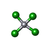

| #2: Chemical | ChemComp-GOL /  Mass: 92.094 Da / Num. of mol.: 16 / Source method: obtained synthetically / Formula: C3H8O3 Mass: 92.094 Da / Num. of mol.: 16 / Source method: obtained synthetically / Formula: C3H8O3#3: Chemical | ChemComp-P3C /  Mass: 301.437 Da / Num. of mol.: 4 / Source method: obtained synthetically / Formula: Cl3Pt Mass: 301.437 Da / Num. of mol.: 4 / Source method: obtained synthetically / Formula: Cl3Pt#4: Chemical | ChemComp-K /  Mass: 39.098 Da / Num. of mol.: 8 / Source method: obtained synthetically / Formula: K Mass: 39.098 Da / Num. of mol.: 8 / Source method: obtained synthetically / Formula: K#5: Chemical | ChemComp-ACT /  Mass: 59.044 Da / Num. of mol.: 13 / Source method: obtained synthetically / Formula: C2H3O2 Mass: 59.044 Da / Num. of mol.: 13 / Source method: obtained synthetically / Formula: C2H3O2#6: Chemical | ChemComp-PC4 /  Mass: 336.890 Da / Num. of mol.: 4 / Source method: obtained synthetically / Formula: Cl4Pt Mass: 336.890 Da / Num. of mol.: 4 / Source method: obtained synthetically / Formula: Cl4Pt#7: Water | ChemComp-HOH / | Mass: 18.015 Da / Num. of mol.: 881 / Source method: isolated from a natural source / Formula: H2O |
|---|
-Details
| Has ligand of interest | N |
|---|
-Experimental details
-Experiment
| Experiment | Method: X-RAY DIFFRACTION / Number of used crystals: 1 |
|---|
- Sample preparation
| Crystal | Density Matthews: 2.2 Å3/Da / Density % sol: 44.18 % |
|---|---|
| Crystal grow | Temperature: 293 K / Method: vapor diffusion, hanging drop Details: 0.2 M ammonium acetate, 0.1 M sodium citrate tribasic dehydrate pH 5.6, 30% polyethylene glycol 4000 |
-Data collection
| Diffraction | Mean temperature: 100 K / Serial crystal experiment: N |
|---|---|
| Diffraction source | Source: SYNCHROTRON / Site: SLS / Beamline: X06DA / Wavelength: 1.0721 Å |
| Detector | Type: MARMOSAIC 225 mm CCD / Detector: CCD / Date: Sep 24, 2009 |
| Radiation | Monochromator: Si111 / Protocol: SINGLE WAVELENGTH / Monochromatic (M) / Laue (L): M / Scattering type: x-ray |
| Radiation wavelength | Wavelength: 1.0721 Å / Relative weight: 1 |
| Reflection | Resolution: 1.35→61.17 Å / Num. obs: 303404 / % possible obs: 98.9 % / Redundancy: 7.4 % / CC1/2: 1 / Net I/σ(I): 17.3 |
| Reflection shell | Resolution: 1.35→1.43 Å / Num. unique obs: 47202 / CC1/2: 0.634 |
-Phasing
| Phasing | Method: SAD |
|---|
- Processing
| Software |
| |||||||||||||||||||||||||||||||||||||||||||||||||||||||||||||||||||||||||||||||||||||||||||||||||||||||||||||||||||||||||||||||||||||||||||||||||||||||||||||||||||||||||||||||||||||||||||||||||||||||||||||||||||||||||
|---|---|---|---|---|---|---|---|---|---|---|---|---|---|---|---|---|---|---|---|---|---|---|---|---|---|---|---|---|---|---|---|---|---|---|---|---|---|---|---|---|---|---|---|---|---|---|---|---|---|---|---|---|---|---|---|---|---|---|---|---|---|---|---|---|---|---|---|---|---|---|---|---|---|---|---|---|---|---|---|---|---|---|---|---|---|---|---|---|---|---|---|---|---|---|---|---|---|---|---|---|---|---|---|---|---|---|---|---|---|---|---|---|---|---|---|---|---|---|---|---|---|---|---|---|---|---|---|---|---|---|---|---|---|---|---|---|---|---|---|---|---|---|---|---|---|---|---|---|---|---|---|---|---|---|---|---|---|---|---|---|---|---|---|---|---|---|---|---|---|---|---|---|---|---|---|---|---|---|---|---|---|---|---|---|---|---|---|---|---|---|---|---|---|---|---|---|---|---|---|---|---|---|---|---|---|---|---|---|---|---|---|---|---|---|---|---|---|---|
| Refinement | Method to determine structure: SAD / Resolution: 1.35→61.17 Å / SU ML: 0.15 / Cross valid method: THROUGHOUT / σ(F): 1.35 / Phase error: 19.01 / Stereochemistry target values: ML
| |||||||||||||||||||||||||||||||||||||||||||||||||||||||||||||||||||||||||||||||||||||||||||||||||||||||||||||||||||||||||||||||||||||||||||||||||||||||||||||||||||||||||||||||||||||||||||||||||||||||||||||||||||||||||
| Solvent computation | Shrinkage radii: 0.9 Å / VDW probe radii: 1.11 Å / Solvent model: FLAT BULK SOLVENT MODEL | |||||||||||||||||||||||||||||||||||||||||||||||||||||||||||||||||||||||||||||||||||||||||||||||||||||||||||||||||||||||||||||||||||||||||||||||||||||||||||||||||||||||||||||||||||||||||||||||||||||||||||||||||||||||||
| Displacement parameters | Biso max: 287.12 Å2 / Biso mean: 20.7761 Å2 / Biso min: 9.32 Å2 | |||||||||||||||||||||||||||||||||||||||||||||||||||||||||||||||||||||||||||||||||||||||||||||||||||||||||||||||||||||||||||||||||||||||||||||||||||||||||||||||||||||||||||||||||||||||||||||||||||||||||||||||||||||||||
| Refinement step | Cycle: final / Resolution: 1.35→61.17 Å
| |||||||||||||||||||||||||||||||||||||||||||||||||||||||||||||||||||||||||||||||||||||||||||||||||||||||||||||||||||||||||||||||||||||||||||||||||||||||||||||||||||||||||||||||||||||||||||||||||||||||||||||||||||||||||
| LS refinement shell | Refine-ID: X-RAY DIFFRACTION / Rfactor Rfree error: 0 / Total num. of bins used: 30
|
