FLUORESCENT PROTEIN / Alternative routes of post-translation chemistry
Function / homology
Function and homology information
host cell membrane / bioluminescence / generation of precursor metabolites and energy / viral envelope / symbiont entry into host cell / virion attachment to host cell / virion membrane / membrane Similarity search - Function
: / Rhabdovirus spike glycoprotein G central domain / Rhabdovirus glycoprotein / Rhabdovirus spike glycoprotein fusion domain / Green fluorescent protein, GFP / Green fluorescent protein-related / Green fluorescent protein / Green fluorescent protein Similarity search - Domain/homology
Mass: 18.015 Da / Num. of mol.: 29 / Source method: isolated from a natural source / Formula: H2O
Has ligand of interest
N
Has protein modification
Y
-
Experimental details
-
Experiment
Experiment
Method: X-RAY DIFFRACTION / Number of used crystals: 1
-
Sample preparation
Crystal
Density Matthews: 2.67 Å3/Da / Density % sol: 53.89 %
Crystal grow
Temperature: 293 K / Method: vapor diffusion, hanging drop Details: 5.7 mg/ml in 20mM Tris 8.0 200 mM NaCl mixed with a 1.44M (NH4)2SO4, 60mM Bicine pH 9.0 reservoir solution.
-
Data collection
Diffraction
Mean temperature: 100 K / Serial crystal experiment: N
Resolution: 2.2→29.4 Å / Cor.coef. Fo:Fc: 0.948 / Cor.coef. Fo:Fc free: 0.895 / SU B: 13.277 / SU ML: 0.306 / SU R Cruickshank DPI: 0.3214 / Cross valid method: THROUGHOUT / σ(F): 0 / ESU R: 0.321 / ESU R Free: 0.287 / Stereochemistry target values: MAXIMUM LIKELIHOOD Details: HYDROGENS HAVE BEEN ADDED IN THE RIDING POSITIONS U VALUES: REFINED INDIVIDUALLY
Rfactor
Num. reflection
% reflection
Selection details
Rfree
0.3396
710
4.9 %
RANDOM
Rwork
0.2543
-
-
-
obs
0.2585
13828
94.83 %
-
Solvent computation
Ion probe radii: 0.8 Å / Shrinkage radii: 0.8 Å / VDW probe radii: 1.2 Å / Solvent model: MASK
In the structure databanks used in Yorodumi, some data are registered as the other names, "COVID-19 virus" and "2019-nCoV". Here are the details of the virus and the list of structure data.
Jan 31, 2019. EMDB accession codes are about to change! (news from PDBe EMDB page)
EMDB accession codes are about to change! (news from PDBe EMDB page)
The allocation of 4 digits for EMDB accession codes will soon come to an end. Whilst these codes will remain in use, new EMDB accession codes will include an additional digit and will expand incrementally as the available range of codes is exhausted. The current 4-digit format prefixed with “EMD-” (i.e. EMD-XXXX) will advance to a 5-digit format (i.e. EMD-XXXXX), and so on. It is currently estimated that the 4-digit codes will be depleted around Spring 2019, at which point the 5-digit format will come into force.
The EM Navigator/Yorodumi systems omit the EMD- prefix.
Related info.:Q: What is EMD? / ID/Accession-code notation in Yorodumi/EM Navigator
Yorodumi is a browser for structure data from EMDB, PDB, SASBDB, etc.
This page is also the successor to EM Navigator detail page, and also detail information page/front-end page for Omokage search.
The word "yorodu" (or yorozu) is an old Japanese word meaning "ten thousand". "mi" (miru) is to see.
Related info.:EMDB / PDB / SASBDB / Comparison of 3 databanks / Yorodumi Search / Aug 31, 2016. New EM Navigator & Yorodumi / Yorodumi Papers / Jmol/JSmol / Function and homology information / Changes in new EM Navigator and Yorodumi
 Movie
Movie Controller


 Open data
 Basic information
 Components
 Keywords
 Function and homology information
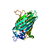
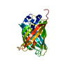
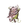
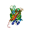
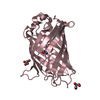
 Recombinant vesicular stomatitis Indiana virus rVSV-G/GFP
 X-RAY DIFFRACTION /
  Authors
 Russian Federation, 1items
  Citation
 Structure visualization
 Downloads & links
 Other downloads


 PDBj
PDBj

 Assembly


 Mass: 96.063 Da / Num. of mol.: 1 / Source method: obtained synthetically / Formula: SO4
 Mass: 18.015 Da / Num. of mol.: 29 / Source method: isolated from a natural source / Formula: H2O
 Sample preparation
 / Beamline: 22-ID / Wavelength: 1 Å
 Processing