 Movie
Movie Controller

[English] 日本語
 Yorodumi
- PDB-6zm3: The structure of an E2 ubiquitin-conjugating complex (UBC2-UEV1) ... -
+ Open data

- Basic information
| Entry | Database: PDB / ID: 6zm3 | |||||||||||||||
|---|---|---|---|---|---|---|---|---|---|---|---|---|---|---|---|---|
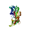
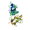
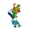
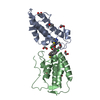
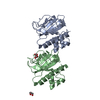
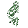
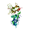
| Title | The structure of an E2 ubiquitin-conjugating complex (UBC2-UEV1) essential for Leishmania amastigote differentiation | |||||||||||||||
 Components Components |
| |||||||||||||||
 Keywords Keywords | SIGNALING PROTEIN / Leishmaniasis / Differentiation / Ubiquitin / Conjugation / UBC2-UEV1 complex | |||||||||||||||
| Function / homology |  Function and homology information Function and homology informationubiquitin-protein ligase / ligase activity / transferase activity / ATP binding Similarity search - Function | |||||||||||||||
| Biological species |   Leishmania mexicana (eukaryote) Leishmania mexicana (eukaryote) | |||||||||||||||
| Method |  X-RAY DIFFRACTION / SYNCHROTRON / MOLECULAR REPLACEMENT / Resolution: 1.7 Å X-RAY DIFFRACTION / SYNCHROTRON / MOLECULAR REPLACEMENT / Resolution: 1.7 Å | |||||||||||||||
 Authors Authors | Burge, R.J. / Dodson, E.J. / Wilkinson, A.J. / Mottram, J.C. | |||||||||||||||
| Funding support |  United Kingdom, 4items United Kingdom, 4items
| |||||||||||||||
 Citation Citation | Journal: Plos Pathog. / Year: 2020 Title: Leishmania differentiation requires ubiquitin conjugation mediated by a UBC2-UEV1 E2 complex. Authors: Burge, R.J. / Damianou, A. / Wilkinson, A.J. / Rodenko, B. / Mottram, J.C. | |||||||||||||||
| History |
|
- Structure visualization
| Structure viewer | Molecule: MolmilJmol/JSmol |
|---|
- Downloads & links
-Download
| PDBx/mmCIF format | 6zm3.cif.gz | 136.8 KB | Display | PDBx/mmCIF format |
|---|---|---|---|---|
| PDB format | pdb6zm3.ent.gz | Display | PDB format | |
| PDBx/mmJSON format | 6zm3.json.gz | Tree view | PDBx/mmJSON format | |
| Others |  Other downloads Other downloads |
-Validation report
| Arichive directory | https://data.pdbj.org/pub/pdb/validation_reports/zm/6zm3ftp://data.pdbj.org/pub/pdb/validation_reports/zm/6zm3 | HTTPS FTP |
|---|
-Related structure data
| Related structure data |  1j7dS S: Starting model for refinement |
|---|---|
| Similar structure data |
-Links
 PDBj
PDBj

- Assembly
| Deposited unit | 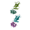
| ||||||||
|---|---|---|---|---|---|---|---|---|---|
| 1 | 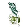
| ||||||||
| 2 | 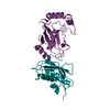
| ||||||||
| Unit cell |
|
-Components
| #1: Protein | Mass: 17298.100 Da / Num. of mol.: 2 Source method: isolated from a genetically manipulated source Source: (gene. exp.) Leishmania mexicana (strain MHOM/GT/2001/U1103) (eukaryote)Strain: MHOM/GT/2001/U1103 / Gene: LMXM_04_0680 / Production host:  #2: Protein | Mass: 16279.417 Da / Num. of mol.: 2 Source method: isolated from a genetically manipulated source Source: (gene. exp.) Leishmania mexicana (strain MHOM/GT/2001/U1103) (eukaryote)Strain: MHOM/GT/2001/U1103 / Gene: LMXM_13_1580 / Production host: #3: Water | ChemComp-HOH / |  Mass: 18.015 Da / Num. of mol.: 267 / Source method: isolated from a natural source / Formula: H2O Mass: 18.015 Da / Num. of mol.: 267 / Source method: isolated from a natural source / Formula: H2OHas ligand of interest | N | |
|---|
-Experimental details
-Experiment
| Experiment | Method: X-RAY DIFFRACTION / Number of used crystals: 1 |
|---|
- Sample preparation
| Crystal | Density Matthews: 2.16 Å3/Da / Density % sol: 44 % |
|---|---|
| Crystal grow | Temperature: 298 K / Method: vapor diffusion, sitting drop / pH: 7.5 Details: UBC2 and UEV1 were mixed in a 1:1 molar ratio to a final concentration of 6.6 mg mL-1 and incubated on ice for 30 min. Crystals were grown using a sitting drop method with a 1:1 ratio of ...Details: UBC2 and UEV1 were mixed in a 1:1 molar ratio to a final concentration of 6.6 mg mL-1 and incubated on ice for 30 min. Crystals were grown using a sitting drop method with a 1:1 ratio of protein to reservoir solution (0.1 M Bis-Tris propane, pH 7.5, 0.2 M sodium formate and 20% PEG) in the drop. Crystals took 2 days to appear. |
-Data collection
| Diffraction | Mean temperature: 100 K / Serial crystal experiment: N | ||||||||||||||||||
|---|---|---|---|---|---|---|---|---|---|---|---|---|---|---|---|---|---|---|---|
| Diffraction source | Source: SYNCHROTRON / Site: Diamond / Beamline: I03 / Wavelength: 0.976254 Å | ||||||||||||||||||
| Detector | Type: DECTRIS EIGER2 XE 16M / Detector: PIXEL / Date: Apr 8, 2019 | ||||||||||||||||||
| Radiation | Protocol: SINGLE WAVELENGTH / Monochromatic (M) / Laue (L): M / Scattering type: x-ray | ||||||||||||||||||
| Radiation wavelength | Wavelength: 0.976254 Å / Relative weight: 1 | ||||||||||||||||||
| Reflection | Resolution: 1.65→47.25 Å / Num. obs: 67149 / % possible obs: 100 % / Redundancy: 4.2 % / CC1/2: 0.996 / Rmerge(I) obs: 0.092 / Rpim(I) all: 0.075 / Rrim(I) all: 0.12 / Net I/σ(I): 6.7 | ||||||||||||||||||
| Reflection shell | Diffraction-ID: 1 / Redundancy: 4.2 %
|
- Processing
| Software |
| |||||||||||||||||||||||||||||||||||||||||||||||||||||||||||||||||||||||||||||||||||||||||||||||||||||||||||||||||||||||||||||||||||||||||||||||||||
|---|---|---|---|---|---|---|---|---|---|---|---|---|---|---|---|---|---|---|---|---|---|---|---|---|---|---|---|---|---|---|---|---|---|---|---|---|---|---|---|---|---|---|---|---|---|---|---|---|---|---|---|---|---|---|---|---|---|---|---|---|---|---|---|---|---|---|---|---|---|---|---|---|---|---|---|---|---|---|---|---|---|---|---|---|---|---|---|---|---|---|---|---|---|---|---|---|---|---|---|---|---|---|---|---|---|---|---|---|---|---|---|---|---|---|---|---|---|---|---|---|---|---|---|---|---|---|---|---|---|---|---|---|---|---|---|---|---|---|---|---|---|---|---|---|---|---|---|---|
| Refinement | Method to determine structure: MOLECULAR REPLACEMENT Starting model: PDB ID: 1J7D Resolution: 1.7→46.25 Å / Cor.coef. Fo:Fc: 0.957 / Cor.coef. Fo:Fc free: 0.946 / SU B: 3.71 / SU ML: 0.117 / Cross valid method: FREE R-VALUE / ESU R: 0.137 / ESU R Free: 0.131
| |||||||||||||||||||||||||||||||||||||||||||||||||||||||||||||||||||||||||||||||||||||||||||||||||||||||||||||||||||||||||||||||||||||||||||||||||||
| Solvent computation | Ion probe radii: 0.8 Å / Shrinkage radii: 0.8 Å / VDW probe radii: 1.2 Å / Solvent model: MASK BULK SOLVENT | |||||||||||||||||||||||||||||||||||||||||||||||||||||||||||||||||||||||||||||||||||||||||||||||||||||||||||||||||||||||||||||||||||||||||||||||||||
| Displacement parameters | Biso mean: 31.043 Å2
| |||||||||||||||||||||||||||||||||||||||||||||||||||||||||||||||||||||||||||||||||||||||||||||||||||||||||||||||||||||||||||||||||||||||||||||||||||
| Refinement step | Cycle: LAST / Resolution: 1.7→46.25 Å
| |||||||||||||||||||||||||||||||||||||||||||||||||||||||||||||||||||||||||||||||||||||||||||||||||||||||||||||||||||||||||||||||||||||||||||||||||||
| Refine LS restraints |
| |||||||||||||||||||||||||||||||||||||||||||||||||||||||||||||||||||||||||||||||||||||||||||||||||||||||||||||||||||||||||||||||||||||||||||||||||||
| LS refinement shell |
|
