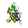
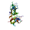
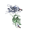
Entry Database : PDB / ID : 2orxTitle Structural Basis for Ligand Binding and Heparin Mediated Activation of Neuropilin Neuropilin-1 Keywords / / / / Function / homology Function Domain/homology Component
/ / / / / / / / / / / / / / / / / / / / / / / / / / / / / / / / / / / / / / / / / / / / / / / / / / / / / / / / / / / / / / / / / / / / / / / / / / / / / / / / / / / / / / / / / / / / / / / / / / / / / / / / / / / / / / / / / / / / / / / / / / / / / / / / / / / / / / / / / / / / / / Biological species Rattus norvegicus (Norway rat)Method / / / Resolution : 2.4 Å Authors Vander Kooi, C.W. / Jusino, M.A. / Perman, B. / Neau, D.B. / Bellamy, H.D. / Leahy, D.J. Journal : Proc.Natl.Acad.Sci.Usa / Year : 2007Title : Structural basis for ligand and heparin binding to neuropilin B domainsAuthors : Vander Kooi, C.W. / Jusino, M.A. / Perman, B. / Neau, D.B. / Bellamy, H.D. / Leahy, D.J. History Deposition Feb 5, 2007 Deposition site / Processing site Revision 1.0 Apr 3, 2007 Provider / Type Revision 1.1 Apr 21, 2008 Group Revision 1.2 Jul 13, 2011 Group / Version format complianceRevision 1.3 Aug 30, 2023 Group / Database references / Refinement descriptionCategory chem_comp_atom / chem_comp_bond ... chem_comp_atom / chem_comp_bond / database_2 / pdbx_initial_refinement_model Item / _database_2.pdbx_database_accessionRevision 1.4 Oct 30, 2024 Group / Category / pdbx_modification_feature
Show all Show less
 Movie
Movie Controller

 Yorodumi
 Open data
 Basic information
 Components
 Keywords
 Function and homology information
 X-RAY DIFFRACTION /
  Authors
 Citation
 Structure visualization
 Downloads & links
 Other downloads
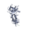


 PDBj
PDBj

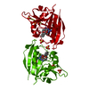
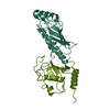
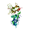
 Assembly
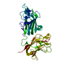

 Mass: 18.015 Da / Num. of mol.: 198 / Source method: isolated from a natural source / Formula: H2O
 Sample preparation
 / Beamline: GCPCC / Wavelength: 1.38 Å
 Processing