 Movie
Movie Controller

[English] 日本語
 Yorodumi
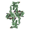
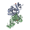
- PDB-7bvv: Crystal structure of sulfonic peroxiredoxin Ahp1 in complex with ... -
+ Open data

- Basic information
| Entry | Database: PDB / ID: 7bvv | ||||||
|---|---|---|---|---|---|---|---|
| Title | Crystal structure of sulfonic peroxiredoxin Ahp1 in complex with thioredoxin Trx2 | ||||||
 Components Components |
| ||||||
 Keywords Keywords | OXIDOREDUCTASE / peroxiredoxin / thioredoxin / alkyl hydroperoxide reductase / complex | ||||||
| Function / homology |  Function and homology information Function and homology informationmembrane fusion priming complex / TP53 Regulates Metabolic Genes / Interconversion of nucleotide di- and triphosphates / The NLRP3 inflammasome / vacuole inheritance / vacuole fusion, non-autophagic / sulfate assimilation / Detoxification of Reactive Oxygen Species / disulfide oxidoreductase activity / thioredoxin-dependent peroxiredoxin ...membrane fusion priming complex / TP53 Regulates Metabolic Genes / Interconversion of nucleotide di- and triphosphates / The NLRP3 inflammasome / vacuole inheritance / vacuole fusion, non-autophagic / sulfate assimilation / Detoxification of Reactive Oxygen Species / disulfide oxidoreductase activity / thioredoxin-dependent peroxiredoxin / fungal-type vacuole / thioredoxin peroxidase activity / retrograde vesicle-mediated transport, Golgi to endoplasmic reticulum / Oxidative Stress Induced Senescence / deoxyribonucleotide biosynthetic process / response to metal ion / protein-disulfide reductase activity / endoplasmic reticulum to Golgi vesicle-mediated transport / cell redox homeostasis / hydrogen peroxide catabolic process / glutathione metabolic process / peroxisome / protein transport / cellular response to oxidative stress / Golgi membrane / mitochondrion / nucleus / plasma membrane / cytoplasm / cytosol Similarity search - Function | ||||||
| Biological species |  | ||||||
| Method |  X-RAY DIFFRACTION / SYNCHROTRON / MOLECULAR REPLACEMENT / Resolution: 2.12 Å X-RAY DIFFRACTION / SYNCHROTRON / MOLECULAR REPLACEMENT / Resolution: 2.12 Å | ||||||
 Authors Authors | Lian, F.M. / Jiang, Y.L. / Yang, W. / Yang, X. | ||||||
 Citation Citation | Journal: Int.J.Biol.Macromol. / Year: 2020 Title: Crystal structure of sulfonic peroxiredoxin Ahp1 in complex with thioredoxin Trx2 mimics a conformational intermediate during the catalytic cycle. Authors: Lian, F.M. / Jiang, Y.L. / Yang, W. / Yang, X. | ||||||
| History |
|
- Structure visualization
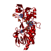
| Structure viewer | Molecule: MolmilJmol/JSmol |
|---|
- Downloads & links
-Download
| PDBx/mmCIF format | 7bvv.cif.gz | 71 KB | Display | PDBx/mmCIF format |
|---|---|---|---|---|
| PDB format | pdb7bvv.ent.gz | 50.2 KB | Display | PDB format |
| PDBx/mmJSON format | 7bvv.json.gz | Tree view | PDBx/mmJSON format | |
| Others |  Other downloads Other downloads |
-Validation report
| Arichive directory | https://data.pdbj.org/pub/pdb/validation_reports/bv/7bvvftp://data.pdbj.org/pub/pdb/validation_reports/bv/7bvv | HTTPS FTP |
|---|
-Related structure data
| Related structure data |  4dssS S: Starting model for refinement |
|---|---|
| Similar structure data |


-Links
 PDBj
PDBj


- Assembly
| Deposited unit | 
| ||||||||
|---|---|---|---|---|---|---|---|---|---|
| 1 | 
| ||||||||
| Unit cell |
| ||||||||
| Components on special symmetry positions |
|
-Components
| #1: Protein | Mass: 19180.598 Da / Num. of mol.: 1 Source method: isolated from a genetically manipulated source Source: (gene. exp.) Strain: ATCC 204508 / S288c / Gene: AHP1, YLR109W, L2916, L9354.5 / Production host:  |
|---|---|
| #2: Protein | Mass: 12214.898 Da / Num. of mol.: 1 / Mutation: C34S Source method: isolated from a genetically manipulated source Source: (gene. exp.) Strain: ATCC 204508 / S288c / Gene: TRX2, TRX1, YGR209C, G7746 / Production host: |
| #3: Water | ChemComp-HOH /  Mass: 18.015 Da / Num. of mol.: 81 / Source method: isolated from a natural source / Formula: H2O Mass: 18.015 Da / Num. of mol.: 81 / Source method: isolated from a natural source / Formula: H2O |
| Has ligand of interest | Y |
-Experimental details
-Experiment
| Experiment | Method: X-RAY DIFFRACTION / Number of used crystals: 1 |
|---|
- Sample preparation
| Crystal | Density Matthews: 2.31 Å3/Da / Density % sol: 46.72 % |
|---|---|
| Crystal grow | Temperature: 289 K / Method: vapor diffusion, sitting drop / pH: 7.5 Details: 25% polyethylene glycol 3,350, 0.2 M lithium sulfate, 0.1 M HEPES-NaOH, pH 7.5 |
-Data collection
| Diffraction | Mean temperature: 100 K / Serial crystal experiment: N |
|---|---|
| Diffraction source | Source: SYNCHROTRON / Site: SSRF  / Beamline: BL17U / Wavelength: 0.9792 Å / Beamline: BL17U / Wavelength: 0.9792 Å |
| Detector | Type: ADSC QUANTUM 315r / Detector: CCD / Date: Nov 13, 2011 |
| Radiation | Protocol: SINGLE WAVELENGTH / Monochromatic (M) / Laue (L): M / Scattering type: x-ray |
| Radiation wavelength | Wavelength: 0.9792 Å / Relative weight: 1 |
| Reflection | Resolution: 2.12→50 Å / Num. obs: 16850 / % possible obs: 100 % / Redundancy: 7.1 % / Biso Wilson estimate: 30.1 Å2 / CC1/2: 0.998 / Rmerge(I) obs: 0.087 / Rpim(I) all: 0.035 / Rrim(I) all: 0.094 / Net I/σ(I): 14.8 |
| Reflection shell | Resolution: 2.12→2.23 Å / Redundancy: 7.3 % / Rmerge(I) obs: 0.493 / Mean I/σ(I) obs: 4.2 / Num. unique obs: 2424 / CC1/2: 0.904 / Rpim(I) all: 0.195 / Rrim(I) all: 0.53 / % possible all: 100 |
- Processing
| Software |
| |||||||||||||||||||||||||||||||||||||||||||||||||||||||||||||||||||||||||||||||||||||||||||||||||||||||||||||||||||||||||||||||||||||||||||||||||||||||||||
|---|---|---|---|---|---|---|---|---|---|---|---|---|---|---|---|---|---|---|---|---|---|---|---|---|---|---|---|---|---|---|---|---|---|---|---|---|---|---|---|---|---|---|---|---|---|---|---|---|---|---|---|---|---|---|---|---|---|---|---|---|---|---|---|---|---|---|---|---|---|---|---|---|---|---|---|---|---|---|---|---|---|---|---|---|---|---|---|---|---|---|---|---|---|---|---|---|---|---|---|---|---|---|---|---|---|---|---|---|---|---|---|---|---|---|---|---|---|---|---|---|---|---|---|---|---|---|---|---|---|---|---|---|---|---|---|---|---|---|---|---|---|---|---|---|---|---|---|---|---|---|---|---|---|---|---|---|
| Refinement | Method to determine structure: MOLECULAR REPLACEMENT Starting model: 4DSS Resolution: 2.12→38.525 Å / Cor.coef. Fo:Fc: 0.942 / Cor.coef. Fo:Fc free: 0.921 / Cross valid method: THROUGHOUT / ESU R: 0.252 / ESU R Free: 0.195 Details: Hydrogens have been added in their riding positions
| |||||||||||||||||||||||||||||||||||||||||||||||||||||||||||||||||||||||||||||||||||||||||||||||||||||||||||||||||||||||||||||||||||||||||||||||||||||||||||
| Solvent computation | Ion probe radii: 0.8 Å / Shrinkage radii: 0.8 Å / VDW probe radii: 1.2 Å | |||||||||||||||||||||||||||||||||||||||||||||||||||||||||||||||||||||||||||||||||||||||||||||||||||||||||||||||||||||||||||||||||||||||||||||||||||||||||||
| Displacement parameters | Biso mean: 33.033 Å2
| |||||||||||||||||||||||||||||||||||||||||||||||||||||||||||||||||||||||||||||||||||||||||||||||||||||||||||||||||||||||||||||||||||||||||||||||||||||||||||
| Refinement step | Cycle: LAST / Resolution: 2.12→38.525 Å
| |||||||||||||||||||||||||||||||||||||||||||||||||||||||||||||||||||||||||||||||||||||||||||||||||||||||||||||||||||||||||||||||||||||||||||||||||||||||||||
| Refine LS restraints |
| |||||||||||||||||||||||||||||||||||||||||||||||||||||||||||||||||||||||||||||||||||||||||||||||||||||||||||||||||||||||||||||||||||||||||||||||||||||||||||
| LS refinement shell |
|
