 Movie
Movie Controller


+ Open data

- Basic information
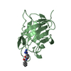
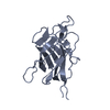
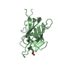
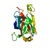
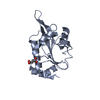
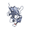
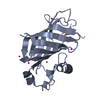
| Entry | Database: PDB / ID: 1kex | ||||||
|---|---|---|---|---|---|---|---|
| Title | Crystal Structure of the b1 Domain of Human Neuropilin-1 | ||||||
 Components Components | Neuropilin-1 | ||||||
 Keywords Keywords | PROTEIN BINDING / beta barrel / jelly-roll | ||||||
| Function / homology |  Function and homology information Function and homology informationendothelial tip cell fate specification / basal dendrite development / otic placode development / basal dendrite arborization / retina vasculature morphogenesis in camera-type eye / dichotomous subdivision of terminal units involved in salivary gland branching / vestibulocochlear nerve structural organization / dorsal root ganglion morphogenesis / ventral trunk neural crest cell migration / sympathetic neuron projection guidance ...endothelial tip cell fate specification / basal dendrite development / otic placode development / basal dendrite arborization / retina vasculature morphogenesis in camera-type eye / dichotomous subdivision of terminal units involved in salivary gland branching / vestibulocochlear nerve structural organization / dorsal root ganglion morphogenesis / ventral trunk neural crest cell migration / sympathetic neuron projection guidance / facioacoustic ganglion development / trigeminal ganglion development / trigeminal nerve structural organization / sensory neuron axon guidance / facial nerve structural organization / negative regulation of axon extension involved in axon guidance / branchiomotor neuron axon guidance / gonadotrophin-releasing hormone neuronal migration to the hypothalamus / protein localization to early endosome / VEGF-activated neuropilin signaling pathway / axon extension involved in axon guidance / renal artery morphogenesis / neurofilament / sympathetic neuron projection extension / Neuropilin interactions with VEGF and VEGFR / vascular endothelial growth factor binding / motor neuron migration / regulation of vascular endothelial growth factor receptor signaling pathway / angiogenesis involved in coronary vascular morphogenesis / postsynapse organization / neural crest cell migration involved in autonomic nervous system development / sympathetic ganglion development / axonogenesis involved in innervation / vascular endothelial growth factor receptor activity / CHL1 interactions / positive regulation of axon extension involved in axon guidance / endothelial cell chemotaxis / regulation of vesicle-mediated transport / semaphorin receptor complex / neuropilin signaling pathway / SEMA3A-Plexin repulsion signaling by inhibiting Integrin adhesion / Signaling by ROBO receptors / substrate-dependent cell migration, cell extension / coronary artery morphogenesis / CRMPs in Sema3A signaling / semaphorin receptor activity / commissural neuron axon guidance / cell migration involved in sprouting angiogenesis / outflow tract septum morphogenesis / axonal fasciculation / motor neuron axon guidance / hepatocyte growth factor receptor signaling pathway / sprouting angiogenesis / regulation of Cdc42 protein signal transduction / retinal ganglion cell axon guidance / artery morphogenesis / positive regulation of filopodium assembly / positive regulation of cell migration involved in sprouting angiogenesis / positive regulation of smooth muscle cell migration / cellular response to hepatocyte growth factor stimulus / neural crest cell migration / branching involved in blood vessel morphogenesis / growth factor binding / positive chemotaxis / sorting endosome / platelet-derived growth factor receptor signaling pathway / cytokine binding / semaphorin-plexin signaling pathway / Sema3A PAK dependent Axon repulsion / cellular response to vascular endothelial growth factor stimulus / positive regulation of phosphorylation / positive regulation of focal adhesion assembly / vascular endothelial growth factor receptor signaling pathway / vasculogenesis / coreceptor activity / positive regulation of stress fiber assembly / positive regulation of endothelial cell proliferation / positive regulation of substrate adhesion-dependent cell spreading / positive regulation of endothelial cell migration / axon guidance / animal organ morphogenesis / negative regulation of extrinsic apoptotic signaling pathway / Signal transduction by L1 / GTPase activator activity / integrin-mediated signaling pathway / response to wounding / mitochondrial membrane / neuron migration / positive regulation of angiogenesis / cell-cell signaling / heparin binding / angiogenesis / cytoplasmic vesicle / negative regulation of neuron apoptotic process / Attachment and Entry / early endosome / positive regulation of ERK1 and ERK2 cascade / postsynaptic membrane / signaling receptor complex / neuron projection Similarity search - Function | ||||||
| Biological species |  Homo sapiens (human) Homo sapiens (human) | ||||||
| Method |  X-RAY DIFFRACTION / MOLECULAR REPLACEMENT / Resolution: 1.9 Å X-RAY DIFFRACTION / MOLECULAR REPLACEMENT / Resolution: 1.9 Å | ||||||
 Authors Authors | Lee, C.C. / Kreusch, A. / McMullan, D. / Ng, K. / Spraggon, G. | ||||||
 Citation Citation | Journal: Structure / Year: 2003 Title: Crystal Structure of the Human Neuropilin-1 b1 Domain Authors: Lee, C.C. / Kreusch, A. / McMullan, D. / Ng, K. / Spraggon, G. | ||||||
| History |
|
- Structure visualization
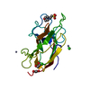
| Structure viewer | Molecule: MolmilJmol/JSmol |
|---|
- Downloads & links
-Download
| PDBx/mmCIF format | 1kex.cif.gz | 47.2 KB | Display | PDBx/mmCIF format |
|---|---|---|---|---|
| PDB format | pdb1kex.ent.gz | 33.3 KB | Display | PDB format |
| PDBx/mmJSON format | 1kex.json.gz | Tree view | PDBx/mmJSON format | |
| Others |  Other downloads Other downloads |
-Validation report
| Arichive directory | https://data.pdbj.org/pub/pdb/validation_reports/ke/1kexftp://data.pdbj.org/pub/pdb/validation_reports/ke/1kex | HTTPS FTP |
|---|
-Related structure data
| Related structure data |  1cztS S: Starting model for refinement |
|---|---|
| Similar structure data |


-Links
 PDBj
PDBj


- Assembly
| Deposited unit | 
| ||||||||
|---|---|---|---|---|---|---|---|---|---|
| 1 |
| ||||||||
| Unit cell |
|
-Components
| #1: Protein | Mass: 17590.896 Da / Num. of mol.: 1 / Fragment: b1 domain Source method: isolated from a genetically manipulated source Source: (gene. exp.) Homo sapiens (human) / Gene: nrp1 / Production host:  Trichoplusia ni (cabbage looper) / References: UniProt: O14786 Trichoplusia ni (cabbage looper) / References: UniProt: O14786 |
|---|---|
| #2: Water | ChemComp-HOH /  Mass: 18.015 Da / Num. of mol.: 201 / Source method: isolated from a natural source / Formula: H2O Mass: 18.015 Da / Num. of mol.: 201 / Source method: isolated from a natural source / Formula: H2O |
| Has protein modification | Y |
-Experimental details
-Experiment
| Experiment | Method: X-RAY DIFFRACTION / Number of used crystals: 1 |
|---|
- Sample preparation
| Crystal | Density Matthews: 2.48 Å3/Da / Density % sol: 50.36 % | ||||||||||||||||||||||||||||||||||||||||||||||||||||||||
|---|---|---|---|---|---|---|---|---|---|---|---|---|---|---|---|---|---|---|---|---|---|---|---|---|---|---|---|---|---|---|---|---|---|---|---|---|---|---|---|---|---|---|---|---|---|---|---|---|---|---|---|---|---|---|---|---|---|
| Crystal grow | Temperature: 277.15 K / Method: vapor diffusion, hanging drop / pH: 6 Details: PEG 8000, MES, zinc acetate, pH 6.0, VAPOR DIFFUSION, HANGING DROP, temperature 277.15K | ||||||||||||||||||||||||||||||||||||||||||||||||||||||||
| Crystal grow | *PLUS Temperature: 4 ℃ / pH: 7.9 | ||||||||||||||||||||||||||||||||||||||||||||||||||||||||
| Components of the solutions | *PLUS
|
-Data collection
| Diffraction source | Source: ROTATING ANODE / Type: RIGAKU FR-D / Wavelength: 1.5418 |
|---|---|
| Detector | Type: RIGAKU / Detector: IMAGE PLATE / Date: Aug 15, 2001 |
| Radiation | Protocol: SINGLE WAVELENGTH / Monochromatic (M) / Laue (L): M / Scattering type: x-ray |
| Radiation wavelength | Wavelength: 1.5418 Å / Relative weight: 1 |
| Reflection | Resolution: 1.9→50 Å / Num. all: 14523 / Num. obs: 12290 / % possible obs: 97.3 % / Rsym value: 0.042 / Net I/σ(I): 32.1 |
| Reflection shell | Resolution: 1.9→2.03 Å / Rsym value: 0.105 / % possible all: 90.5 |
| Reflection | *PLUS Num. measured all: 370706 / Rmerge(I) obs: 0.042 |
| Reflection shell | *PLUS % possible obs: 90.5 % / Rmerge(I) obs: 0.105 |
- Processing
| Software |
| ||||||||||||||||||||||||||||||||||||||||||||||||||||||||||||
|---|---|---|---|---|---|---|---|---|---|---|---|---|---|---|---|---|---|---|---|---|---|---|---|---|---|---|---|---|---|---|---|---|---|---|---|---|---|---|---|---|---|---|---|---|---|---|---|---|---|---|---|---|---|---|---|---|---|---|---|---|---|
| Refinement | Method to determine structure: MOLECULAR REPLACEMENT Starting model: PDB ENTRY 1CZT Resolution: 1.9→500 Å /
| ||||||||||||||||||||||||||||||||||||||||||||||||||||||||||||
| Refinement step | Cycle: LAST / Resolution: 1.9→500 Å
| ||||||||||||||||||||||||||||||||||||||||||||||||||||||||||||
| Refine LS restraints |
| ||||||||||||||||||||||||||||||||||||||||||||||||||||||||||||
| Refinement | *PLUS Lowest resolution: 50 Å | ||||||||||||||||||||||||||||||||||||||||||||||||||||||||||||
| Solvent computation | *PLUS | ||||||||||||||||||||||||||||||||||||||||||||||||||||||||||||
| Displacement parameters | *PLUS |
