 Movie
Movie Controller

[English] 日本語
 Yorodumi
- PDB-6zll: Crystal Structure of UDP-Glucuronic acid 4-epimerase from Bacillu... -
+ Open data

- Basic information
| Entry | Database: PDB / ID: 6zll | ||||||
|---|---|---|---|---|---|---|---|
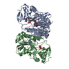
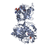
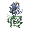
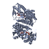
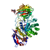
| Title | Crystal Structure of UDP-Glucuronic acid 4-epimerase from Bacillus cereus in complex with UDP-Galacturonic acid and NAD | ||||||
 Components Components | Epimerase domain-containing protein | ||||||
 Keywords Keywords | OXIDOREDUCTASE / epimerase / UDP-Galacturonic acid / NAD / UDP-sugar binding protein | ||||||
| Function / homology | NAD-dependent epimerase/dehydratase / NAD dependent epimerase/dehydratase family / catalytic activity / NAD(P)-binding domain superfamily / NICOTINAMIDE-ADENINE-DINUCLEOTIDE / Chem-UGB / Epimerase domain-containing protein Function and homology information Function and homology information | ||||||
| Biological species |  | ||||||
| Method |  X-RAY DIFFRACTION / SYNCHROTRON / MOLECULAR REPLACEMENT / Resolution: 1.85 Å X-RAY DIFFRACTION / SYNCHROTRON / MOLECULAR REPLACEMENT / Resolution: 1.85 Å | ||||||
 Authors Authors | Iacovino, L.G. / Savino, S. / Mattevi, A. | ||||||
| Funding support |  Italy, 1items Italy, 1items
| ||||||
 Citation Citation | Journal: J.Biol.Chem. / Year: 2020 Title: Crystallographic snapshots of UDP-glucuronic acid 4-epimerase ligand binding, rotation, and reduction. Authors: Iacovino, L.G. / Savino, S. / Borg, A.J.E. / Binda, C. / Nidetzky, B. / Mattevi, A. | ||||||
| History |
|
- Structure visualization
| Structure viewer | Molecule: MolmilJmol/JSmol |
|---|
- Downloads & links
-Download
| PDBx/mmCIF format | 6zll.cif.gz | 268.7 KB | Display | PDBx/mmCIF format |
|---|---|---|---|---|
| PDB format | pdb6zll.ent.gz | 216.4 KB | Display | PDB format |
| PDBx/mmJSON format | 6zll.json.gz | Tree view | PDBx/mmJSON format | |
| Others |  Other downloads Other downloads |
-Validation report
| Arichive directory | https://data.pdbj.org/pub/pdb/validation_reports/zl/6zllftp://data.pdbj.org/pub/pdb/validation_reports/zl/6zll | HTTPS FTP |
|---|
-Related structure data
| Related structure data | 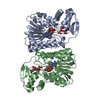 6zl6C 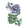 6zlaC 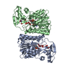 6zldC 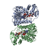 6zljC  6zlkC  5u4qS S: Starting model for refinement C: citing same article ( |
|---|---|
| Similar structure data |


-Links
 PDBj
PDBj- Assembly
| Deposited unit | 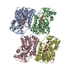
| ||||||||
|---|---|---|---|---|---|---|---|---|---|
| 1 | 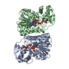
| ||||||||
| 2 | 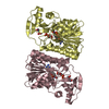
| ||||||||
| Unit cell |
|
-Components
| #1: Protein | Mass: 36526.957 Da / Num. of mol.: 4 Source method: isolated from a genetically manipulated source Source: (gene. exp.) #2: Chemical | ChemComp-UGB / (   Mass: 580.285 Da / Num. of mol.: 4 / Source method: obtained synthetically / Formula: C15H22N2O18P2 / Feature type: SUBJECT OF INVESTIGATION Mass: 580.285 Da / Num. of mol.: 4 / Source method: obtained synthetically / Formula: C15H22N2O18P2 / Feature type: SUBJECT OF INVESTIGATION#3: Chemical | ChemComp-NAD /   Mass: 663.425 Da / Num. of mol.: 4 / Source method: obtained synthetically / Formula: C21H27N7O14P2 / Feature type: SUBJECT OF INVESTIGATION / Comment: NAD*YM Mass: 663.425 Da / Num. of mol.: 4 / Source method: obtained synthetically / Formula: C21H27N7O14P2 / Feature type: SUBJECT OF INVESTIGATION / Comment: NAD*YM#4: Water | ChemComp-HOH / |  Mass: 18.015 Da / Num. of mol.: 243 / Source method: isolated from a natural source / Formula: H2O Mass: 18.015 Da / Num. of mol.: 243 / Source method: isolated from a natural source / Formula: H2OHas ligand of interest | Y | |
|---|
-Experimental details
-Experiment
| Experiment | Method: X-RAY DIFFRACTION / Number of used crystals: 1 |
|---|
- Sample preparation
| Crystal | Density Matthews: 2.53 Å3/Da / Density % sol: 51.43 % |
|---|---|
| Crystal grow | Temperature: 277 K / Method: vapor diffusion / pH: 6.5 Details: 200 mM potassium acetate, 14-24% PEG3350, 2 mM NAD+, 2 mM UDP-Galacturonic acid |
-Data collection
| Diffraction | Mean temperature: 100 K / Serial crystal experiment: N |
|---|---|
| Diffraction source | Source: SYNCHROTRON / Site: SLS  / Beamline: X06SA / Wavelength: 0.98 Å / Beamline: X06SA / Wavelength: 0.98 Å |
| Detector | Type: DECTRIS EIGER X 16M / Detector: PIXEL / Date: Mar 29, 2019 |
| Radiation | Protocol: SINGLE WAVELENGTH / Monochromatic (M) / Laue (L): M / Scattering type: x-ray |
| Radiation wavelength | Wavelength: 0.98 Å / Relative weight: 1 |
| Reflection | Resolution: 1.85→46.09 Å / Num. obs: 107680 / % possible obs: 93 % / Redundancy: 6.9 % / CC1/2: 0.99 / Net I/σ(I): 8.7 |
| Reflection shell | Resolution: 1.85→1.898 Å / Num. unique obs: 7482 / CC1/2: 0.46 |
- Processing
| Software |
| ||||||||||||||||||||||||||||||||||||||||||||||||||||||||||||
|---|---|---|---|---|---|---|---|---|---|---|---|---|---|---|---|---|---|---|---|---|---|---|---|---|---|---|---|---|---|---|---|---|---|---|---|---|---|---|---|---|---|---|---|---|---|---|---|---|---|---|---|---|---|---|---|---|---|---|---|---|---|
| Refinement | Method to determine structure: MOLECULAR REPLACEMENT Starting model: 5U4Q Resolution: 1.85→46.09 Å / Cor.coef. Fo:Fc: 0.968 / Cor.coef. Fo:Fc free: 0.955 / SU B: 4.552 / SU ML: 0.124 / Cross valid method: THROUGHOUT / σ(F): 0 / ESU R: 0.145 / ESU R Free: 0.134 / Stereochemistry target values: MAXIMUM LIKELIHOOD Details: HYDROGENS HAVE BEEN ADDED IN THE RIDING POSITIONS U VALUES : REFINED INDIVIDUALLY
| ||||||||||||||||||||||||||||||||||||||||||||||||||||||||||||
| Solvent computation | Ion probe radii: 0.8 Å / Shrinkage radii: 0.8 Å / VDW probe radii: 1.2 Å / Solvent model: MASK | ||||||||||||||||||||||||||||||||||||||||||||||||||||||||||||
| Displacement parameters | Biso max: 109.18 Å2 / Biso mean: 37.109 Å2 / Biso min: 22.64 Å2
| ||||||||||||||||||||||||||||||||||||||||||||||||||||||||||||
| Refinement step | Cycle: final / Resolution: 1.85→46.09 Å
| ||||||||||||||||||||||||||||||||||||||||||||||||||||||||||||
| Refine LS restraints |
| ||||||||||||||||||||||||||||||||||||||||||||||||||||||||||||
| LS refinement shell | Resolution: 1.85→1.898 Å / Rfactor Rfree error: 0 / Total num. of bins used: 20
|
