 Movie
Movie Controller

[English] 日本語
 Yorodumi
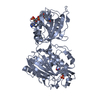
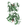
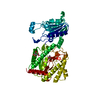
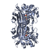
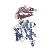
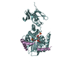
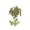
- PDB-3owg: Crystal structure of vaccinia virus Polyadenylate polymerase(vp55) -
+ Open data

- Basic information
| Entry | Database: PDB / ID: 3owg | ||||||
|---|---|---|---|---|---|---|---|
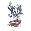
| Title | Crystal structure of vaccinia virus Polyadenylate polymerase(vp55) | ||||||
 Components Components | Poly(A) polymerase catalytic subunit | ||||||
 Keywords Keywords | TRANSFERASE / RNA polyadenylate polymerase complex / translocation / polyadenylate polymerase | ||||||
| Function / homology |  Function and homology information Function and homology informationpolynucleotide adenylyltransferase / poly(A) RNA polymerase activity / mRNA processing / ATP binding / metal ion binding Similarity search - Function | ||||||
| Biological species |  Vaccinia virus WR Vaccinia virus WR | ||||||
| Method |  X-RAY DIFFRACTION / SYNCHROTRON / MOLECULAR REPLACEMENT / Resolution: 2.86 Å X-RAY DIFFRACTION / SYNCHROTRON / MOLECULAR REPLACEMENT / Resolution: 2.86 Å | ||||||
 Authors Authors | Li, C. / Li, H. / Zhou, S. / Gershon, P.D. / Poulos, T.L. | ||||||
 Citation Citation | Journal: Acta Crystallogr.,Sect.D / Year: 2013 Title: Domain-level rocking motion within a polymerase that translocates on single-stranded nucleic acid. Authors: Li, H. / Li, C. / Zhou, S. / Poulos, T.L. / Gershon, P.D. #1: Journal: Mol.Cell / Year: 2006Title: Crystal structures of the vaccinia virus polyadenylate polymerase heterodimer: insights into ATP selectivity and processivity. Authors: Moure, C.M. / Bowman, B.R. / Gershon, P.D. / Quiocho, F.A. #2: Journal: Structure / Year: 2009Title: Polymerase translocation with respect to single-stranded nucleic acid: looping or wrapping of primer around a poly(A) polymerase. Authors: Li, C. / Li, H. / Zhou, S. / Sun, E. / Yoshizawa, J. / Poulos, T.L. / Gershon, P.D. | ||||||
| History |
|
- Structure visualization
| Structure viewer | Molecule: MolmilJmol/JSmol |
|---|
- Downloads & links
-Download
| PDBx/mmCIF format | 3owg.cif.gz | 176.1 KB | Display | PDBx/mmCIF format |
|---|---|---|---|---|
| PDB format | pdb3owg.ent.gz | 142.5 KB | Display | PDB format |
| PDBx/mmJSON format | 3owg.json.gz | Tree view | PDBx/mmJSON format | |
| Others |  Other downloads Other downloads |
-Validation report
| Arichive directory | https://data.pdbj.org/pub/pdb/validation_reports/ow/3owgftp://data.pdbj.org/pub/pdb/validation_reports/ow/3owg | HTTPS FTP |
|---|
-Related structure data
| Related structure data | 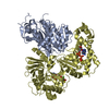 2ga9S S: Starting model for refinement |
|---|---|
| Similar structure data |


-Links
 PDBj
PDBj
- Assembly
| Deposited unit | 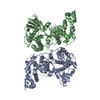
| ||||||||
|---|---|---|---|---|---|---|---|---|---|
| 1 | 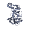
| ||||||||
| 2 | 
| ||||||||
| Unit cell |
|
-Components
| #1: Protein | Mass: 56403.004 Da / Num. of mol.: 2 Source method: isolated from a genetically manipulated source Source: (gene. exp.) Vaccinia virus WR / Strain: Western Reserve / Gene: PAPL, VACWR057, E1L / Plasmid: pPG198 / Production host:  References: UniProt: P23371, polynucleotide adenylyltransferase |
|---|
-Experimental details
-Experiment
| Experiment | Method: X-RAY DIFFRACTION / Number of used crystals: 1 |
|---|
- Sample preparation
| Crystal | Density Matthews: 2.8 Å3/Da / Density % sol: 56.03 % |
|---|---|
| Crystal grow | Temperature: 277 K / Method: vapor diffusion, sitting drop Details: 12-16%PEG 3350,200mM Na-citate, 5% glycero, 1mM DTT, pH ~9, VAPOR DIFFUSION, SITTING DROP, temperature 277K |
-Data collection
| Diffraction | Mean temperature: 110 K |
|---|---|
| Diffraction source | Source: SYNCHROTRON / Site: SSRL  / Beamline: BL9-1 / Wavelength: 0.976 Å / Beamline: BL9-1 / Wavelength: 0.976 Å |
| Detector | Type: ADSC QUANTUM 315 / Detector: CCD / Date: May 3, 2008 / Details: mirrors |
| Radiation | Monochromator: graphite / Protocol: SINGLE WAVELENGTH / Monochromatic (M) / Laue (L): M / Scattering type: x-ray |
| Radiation wavelength | Wavelength: 0.976 Å / Relative weight: 1 |
| Reflection | Resolution: 2.85→50 Å / Num. obs: 29731 / % possible obs: 99.9 % / Observed criterion σ(I): -3 / Redundancy: 3.7 % / Biso Wilson estimate: 90 Å2 / Rmerge(I) obs: 0.06 / Rsym value: 0.06 / Net I/σ(I): 22.6 |
| Reflection shell | Resolution: 2.85→2.9 Å / Redundancy: 3.8 % / Rmerge(I) obs: 0.577 / Mean I/σ(I) obs: 2 / Rsym value: 0.577 / % possible all: 100 |
- Processing
| Software |
| ||||||||||||||||||||
|---|---|---|---|---|---|---|---|---|---|---|---|---|---|---|---|---|---|---|---|---|---|
| Refinement | Method to determine structure: MOLECULAR REPLACEMENT Starting model: PDB entry 2GA9 Resolution: 2.86→41.77 Å / Rfactor Rfree error: 0.008 / Data cutoff high absF: 1227549.9 / Data cutoff low absF: 0 / Isotropic thermal model: GROUP / Cross valid method: THROUGHOUT / σ(F): 0 / Stereochemistry target values: Engh & Huber Details: BULK SOLVENT MODEL USED THE NCS RESTRAINTS (WEIGHT=100)WAS IMPOSED DURING REFINEMENTS
| ||||||||||||||||||||
| Solvent computation | Solvent model: FLAT MODEL / Bsol: 67.8296 Å2 / ksol: 0.3 e/Å3 | ||||||||||||||||||||
| Displacement parameters | Biso mean: 97.9 Å2
| ||||||||||||||||||||
| Refine analyze |
| ||||||||||||||||||||
| Refinement step | Cycle: LAST / Resolution: 2.86→41.77 Å
| ||||||||||||||||||||
| Refine LS restraints |
| ||||||||||||||||||||
| Refine LS restraints NCS | NCS model details: RESTRAINT | ||||||||||||||||||||
| LS refinement shell | Resolution: 2.85→3.03 Å / Rfactor Rfree error: 0.028 / Total num. of bins used: 6
| ||||||||||||||||||||
| Xplor file | Serial no: 1 / Param file: protein_rep.param / Topol file: protein.top |
