Mass: 18.015 Da / Num. of mol.: 701 / Source method: isolated from a natural source / Formula: H2O
Has protein modification
Y
Sequence details
THE CONSTRUCT WAS EXPRESSED WITH A PURIFICATION TAG MGSDKIHHHHHHENLYFQG. THE TAG WAS REMOVED WITH ...THE CONSTRUCT WAS EXPRESSED WITH A PURIFICATION TAG MGSDKIHHHHHHENLYFQG. THE TAG WAS REMOVED WITH TEV PROTEASE LEAVING ONLY A GLYCINE (0) FOLLOWED BY THE TARGET SEQUENCE.
-
Experimental details
-
Experiment
Experiment
Method: X-RAY DIFFRACTION / Number of used crystals: 1
-
Sample preparation
Crystal
Density Matthews: 2.2 Å3/Da / Density % sol: 44.08 %
Crystal grow
Temperature: 277 K / Method: vapor diffusion, sitting drop / pH: 8.6 Details: 0.4000M magnesium chloride, 18.0000% polyethylene glycol 8000, 0.1M TRIS pH 8.6, NANODROP, VAPOR DIFFUSION, SITTING DROP, temperature 277K
Type: MARMOSAIC 325 mm CCD / Detector: CCD / Date: Jun 12, 2009 / Details: Flat mirror (vertical focusing)
Radiation
Monochromator: Single crystal Si(111) bent monochromator (horizontal focusing) Protocol: MAD / Monochromatic (M) / Laue (L): M / Scattering type: x-ray
Radiation wavelength
ID
Wavelength (Å)
Relative weight
1
0.91837
1
2
0.97917
1
3
0.97862
1
Reflection
Resolution: 1.6→27.277 Å / Num. obs: 78581 / % possible obs: 99.3 % / Redundancy: 2.6 % / Biso Wilson estimate: 16.453 Å2 / Rmerge(I) obs: 0.074 / Rsym value: 0.074 / Net I/σ(I): 9.3
Reflection shell
Diffraction-ID: 1
Resolution (Å)
Redundancy (%)
Rmerge(I) obs
Mean I/σ(I) obs
Num. measured all
Num. unique all
Rsym value
% possible all
1.6-1.64
2.5
0.417
2
14633
5758
0.417
98.4
1.64-1.69
2.5
0.371
2.3
14406
5650
0.371
98.6
1.69-1.74
2.5
0.31
2.7
13988
5491
0.31
98.9
1.74-1.79
2.6
0.261
3.4
13511
5297
0.261
99.1
1.79-1.85
2.6
0.208
4.3
13218
5161
0.208
99.2
1.85-1.91
2.6
0.173
5.1
12734
4978
0.173
99.2
1.91-1.98
2.6
0.14
6.4
12396
4842
0.14
99.3
1.98-2.07
2.6
0.114
7.8
11977
4670
0.114
99.3
2.07-2.16
2.6
0.095
9.4
11416
4461
0.095
99.5
2.16-2.26
2.6
0.089
10.3
10984
4285
0.089
99.6
2.26-2.39
2.6
0.079
11.5
10488
4092
0.079
99.7
2.39-2.53
2.6
0.076
12.9
9914
3867
0.076
99.7
2.53-2.7
2.6
0.074
14
9321
3626
0.074
99.9
2.7-2.92
2.6
0.065
16
8657
3366
0.065
99.9
2.92-3.2
2.6
0.055
17.9
7999
3120
0.055
99.9
3.2-3.58
2.6
0.046
20.2
7302
2837
0.046
99.8
3.58-4.13
2.6
0.045
21.5
6362
2462
0.045
99.7
4.13-5.06
2.6
0.046
22.2
5462
2121
0.046
99.4
5.06-7.16
2.6
0.054
21.3
4151
1616
0.054
99.4
7.16-27.28
2.5
0.054
22.6
2240
881
0.054
97.1
-
Phasing
Phasing
Method: MAD
-
Processing
Software
Name
Version
Classification
NB
REFMAC
5.5.0053
refinement
PHENIX
refinement
SHELX
phasing
MolProbity
3beta29
modelbuilding
SCALA
3.2.5
datascaling
PDB_EXTRACT
3.006
dataextraction
MOSFLM
datareduction
SHELXD
phasing
autoSHARP
phasing
Refinement
Method to determine structure: MAD / Resolution: 1.6→27.277 Å / Cor.coef. Fo:Fc: 0.967 / Cor.coef. Fo:Fc free: 0.952 / Occupancy max: 1 / Occupancy min: 0.2 / SU B: 3.369 / SU ML: 0.053 / TLS residual ADP flag: LIKELY RESIDUAL / Cross valid method: THROUGHOUT / σ(F): 0 / ESU R: 0.083 / ESU R Free: 0.083 Stereochemistry target values: MAXIMUM LIKELIHOOD WITH PHASES Details: 1.HYDROGENS HAVE BEEN ADDED IN THE RIDING POSITIONS. 2.ATOM RECORD CONTAINS RESIDUAL B FACTORS ONLY. 3.A MET-INHIBITION PROTOCOL WAS USED FOR SELENOMETHIONINE INCORPORATION DURING PROTEIN ...Details: 1.HYDROGENS HAVE BEEN ADDED IN THE RIDING POSITIONS. 2.ATOM RECORD CONTAINS RESIDUAL B FACTORS ONLY. 3.A MET-INHIBITION PROTOCOL WAS USED FOR SELENOMETHIONINE INCORPORATION DURING PROTEIN EXPRESSION. THE OCCUPANCY OF THE SE ATOMS IN THE MSE RESIDUES WAS REDUCED TO 0.75 TO ACCOUNT FOR THE REDUCED SCATTERING POWER DUE TO PARTIAL S-MET INCORPORATION. 4.MAGNESIUM IONS(MG) AND CHLORIDE IONS(CL) FROM CRYSTALLIZATION ARE MODELED IN THE STRUCTURE. 5.UNKNOWN LIGAND (UNL) IS MODELED NEAR THE MAGNESIUM ION COMPLXED WITH RESIDUES 49, 148 AND 151 IN EACH SUBUNIT, WHICH MAY RESEMBLES GLYCEROL MOLECULE(GOL) MOLECULE.
Rfactor
Num. reflection
% reflection
Selection details
Rfree
0.187
3949
5 %
RANDOM
Rwork
0.156
-
-
-
obs
0.158
78539
99.19 %
-
Solvent computation
Ion probe radii: 0.8 Å / Shrinkage radii: 0.8 Å / VDW probe radii: 1.2 Å / Solvent model: MASK
In the structure databanks used in Yorodumi, some data are registered as the other names, "COVID-19 virus" and "2019-nCoV". Here are the details of the virus and the list of structure data.
Jan 31, 2019. EMDB accession codes are about to change! (news from PDBe EMDB page)
EMDB accession codes are about to change! (news from PDBe EMDB page)
The allocation of 4 digits for EMDB accession codes will soon come to an end. Whilst these codes will remain in use, new EMDB accession codes will include an additional digit and will expand incrementally as the available range of codes is exhausted. The current 4-digit format prefixed with “EMD-” (i.e. EMD-XXXX) will advance to a 5-digit format (i.e. EMD-XXXXX), and so on. It is currently estimated that the 4-digit codes will be depleted around Spring 2019, at which point the 5-digit format will come into force.
The EM Navigator/Yorodumi systems omit the EMD- prefix.
Related info.:Q: What is EMD? / ID/Accession-code notation in Yorodumi/EM Navigator
Yorodumi is a browser for structure data from EMDB, PDB, SASBDB, etc.
This page is also the successor to EM Navigator detail page, and also detail information page/front-end page for Omokage search.
The word "yorodu" (or yorozu) is an old Japanese word meaning "ten thousand". "mi" (miru) is to see.
Related info.:EMDB / PDB / SASBDB / Comparison of 3 databanks / Yorodumi Search / Aug 31, 2016. New EM Navigator & Yorodumi / Yorodumi Papers / Jmol/JSmol / Function and homology information / Changes in new EM Navigator and Yorodumi
 Movie
Movie Controller

 Yorodumi
 Open data
 Basic information
 Components
 Keywords
 Function and homology information
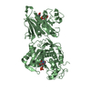
 Silicibacter pomeroyi (bacteria)
 X-RAY DIFFRACTION /
  Authors
 Citation
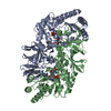
 Structure visualization
 Downloads & links
 Other downloads
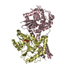


 PDBj
PDBj

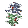
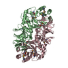
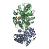
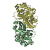
 Assembly
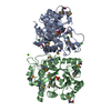

 Mass: 24.305 Da / Num. of mol.: 3 / Source method: obtained synthetically / Formula: Mg
 Mass: 35.453 Da / Num. of mol.: 5 / Source method: obtained synthetically / Formula: Cl
 Mass: 18.015 Da / Num. of mol.: 701 / Source method: isolated from a natural source / Formula: H2O
 Sample preparation
 / Beamline: BL11-1 / Wavelength: 0.91837,0.97917,0.97862
 Processing