 Movie
Movie Controller

[English] 日本語
 Yorodumi
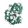
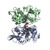
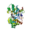
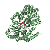
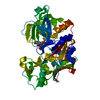
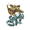
- PDB-6zj7: Trehalose transferase (TreT) from Thermoproteus uzoniensis soaked... -
+ Open data

- Basic information
| Entry | Database: PDB / ID: 6zj7 | ||||||
|---|---|---|---|---|---|---|---|
| Title | Trehalose transferase (TreT) from Thermoproteus uzoniensis soaked with Mg | ||||||
 Components Components | Trehalose phosphorylase/synthase | ||||||
 Keywords Keywords | TRANSFERASE / trehalose transferase / retaining glycosyltransferase / glycosidic bond formation | ||||||
| Function / homology | : / : / Trehalose synthase, N-terminal domain / Glycosyl transferase, family 1 / Glycosyl transferases group 1 / glycosyltransferase activity / glucose metabolic process / THIOCYANATE ION / Trehalose phosphorylase/synthase Function and homology information Function and homology information | ||||||
| Biological species |   Thermoproteus uzoniensis (archaea) Thermoproteus uzoniensis (archaea) | ||||||
| Method |  X-RAY DIFFRACTION / SYNCHROTRON / MOLECULAR REPLACEMENT / Resolution: 2.15 Å X-RAY DIFFRACTION / SYNCHROTRON / MOLECULAR REPLACEMENT / Resolution: 2.15 Å | ||||||
 Authors Authors | Bento, I. / Mestrom, L. / Marsden, S.R. / van der Eijk, H. / Laustsen, J.U. / Jeffries, C.M. / Svergun, D.I. / Hagedoorn, P.-H. / Hanefeld, U. | ||||||
| Funding support |  Germany, 1items Germany, 1items
| ||||||
 Citation Citation | Journal: Acs Catalysis / Year: 2020 Title: Anomeric Selectivity of Trehalose Transferase with Rare l-Sugars. Authors: Mestrom, L. / Marsden, S.R. / van der Eijk, H. / Laustsen, J.U. / Jeffries, C.M. / Svergun, D.I. / Hagedoorn, P.L. / Bento, I. / Hanefeld, U. | ||||||
| History |
|
- Structure visualization
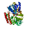
| Structure viewer | Molecule: MolmilJmol/JSmol |
|---|
- Downloads & links
-Download
| PDBx/mmCIF format | 6zj7.cif.gz | 171.2 KB | Display | PDBx/mmCIF format |
|---|---|---|---|---|
| PDB format | pdb6zj7.ent.gz | Display | PDB format | |
| PDBx/mmJSON format | 6zj7.json.gz | Tree view | PDBx/mmJSON format | |
| Others |  Other downloads Other downloads |
-Validation report
| Arichive directory | https://data.pdbj.org/pub/pdb/validation_reports/zj/6zj7ftp://data.pdbj.org/pub/pdb/validation_reports/zj/6zj7 | HTTPS FTP |
|---|
-Related structure data
| Related structure data | 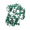 6zj4C  6zjhC 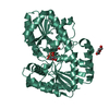 6zmzC 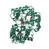 6zn1C C: citing same article ( |
|---|---|
| Similar structure data | |
| Other databases |
|


-Links
 PDBj
PDBj

- Assembly
| Deposited unit | 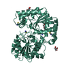
| ||||||||
|---|---|---|---|---|---|---|---|---|---|
| 1 |
| ||||||||
| Unit cell |
|
-Components
| #1: Protein | Mass: 45503.316 Da / Num. of mol.: 1 Source method: isolated from a genetically manipulated source Source: (gene. exp.) Thermoproteus uzoniensis (strain 768-20) (archaea)Strain: 768-20 / Gene: TUZN_0976 / Production host:  | ||||||||
|---|---|---|---|---|---|---|---|---|---|
| #2: Chemical | ChemComp-SCN /   Mass: 58.082 Da / Num. of mol.: 4 / Source method: obtained synthetically / Formula: CNS Mass: 58.082 Da / Num. of mol.: 4 / Source method: obtained synthetically / Formula: CNS#3: Chemical | ChemComp-PG4 / |   Mass: 194.226 Da / Num. of mol.: 1 / Source method: obtained synthetically / Formula: C8H18O5 / Comment: precipitant*YM Mass: 194.226 Da / Num. of mol.: 1 / Source method: obtained synthetically / Formula: C8H18O5 / Comment: precipitant*YM#4: Chemical |   Mass: 92.094 Da / Num. of mol.: 2 / Source method: isolated from a natural source / Formula: C3H8O3 Mass: 92.094 Da / Num. of mol.: 2 / Source method: isolated from a natural source / Formula: C3H8O3#5: Water | ChemComp-HOH / |  Mass: 18.015 Da / Num. of mol.: 88 / Source method: isolated from a natural source / Formula: H2O Mass: 18.015 Da / Num. of mol.: 88 / Source method: isolated from a natural source / Formula: H2OHas ligand of interest | N | |
-Experimental details
-Experiment
| Experiment | Method: X-RAY DIFFRACTION / Number of used crystals: 1 |
|---|
- Sample preparation
| Crystal | Density Matthews: 2.55 Å3/Da / Density % sol: 51.7 % |
|---|---|
| Crystal grow | Temperature: 292 K / Method: vapor diffusion / pH: 6.5 Details: Bis-Tris Propane, PEG 3350, potassium thiocyanate, PEG 400 |
-Data collection
| Diffraction | Mean temperature: 100 K / Serial crystal experiment: N |
|---|---|
| Diffraction source | Source: SYNCHROTRON / Site: PETRA III, EMBL c/o DESY / Beamline: P13 (MX1) / Wavelength: 0.9763 Å |
| Detector | Type: DECTRIS PILATUS 6M-F / Detector: PIXEL / Date: Oct 2, 2019 |
| Radiation | Monochromator: double-crystal monochromator FMB-OXFORD (Oxford, UK) Protocol: SINGLE WAVELENGTH / Monochromatic (M) / Laue (L): M / Scattering type: x-ray |
| Radiation wavelength | Wavelength: 0.9763 Å / Relative weight: 1 |
| Reflection | Resolution: 2.15→110.45 Å / Num. obs: 25646 / % possible obs: 98.8 % / Redundancy: 7.8 % / CC1/2: 0.999 / Rmerge(I) obs: 0.07 / Rpim(I) all: 0.039 / Rrim(I) all: 0.081 / Net I/σ(I): 17.1 |
| Reflection shell | Resolution: 2.15→2.21 Å / Redundancy: 8.1 % / Rmerge(I) obs: 0.024 / Num. unique obs: 2477 / CC1/2: 0.7 / Rpim(I) all: 0.013 / Rrim(I) all: 0.028 |
- Processing
| Software |
| ||||||||||||||||||||||||||||||||||||||||||||||||||||||||||||||||||||||||||||||||||||||||||||||||||||||||||||||||||||||||||||||||||||||||||||||||||||||
|---|---|---|---|---|---|---|---|---|---|---|---|---|---|---|---|---|---|---|---|---|---|---|---|---|---|---|---|---|---|---|---|---|---|---|---|---|---|---|---|---|---|---|---|---|---|---|---|---|---|---|---|---|---|---|---|---|---|---|---|---|---|---|---|---|---|---|---|---|---|---|---|---|---|---|---|---|---|---|---|---|---|---|---|---|---|---|---|---|---|---|---|---|---|---|---|---|---|---|---|---|---|---|---|---|---|---|---|---|---|---|---|---|---|---|---|---|---|---|---|---|---|---|---|---|---|---|---|---|---|---|---|---|---|---|---|---|---|---|---|---|---|---|---|---|---|---|---|---|---|---|---|
| Refinement | Method to determine structure: MOLECULAR REPLACEMENT Starting model: apo-TreT Resolution: 2.15→58.757 Å / Cor.coef. Fo:Fc: 0.961 / Cor.coef. Fo:Fc free: 0.935 / WRfactor Rfree: 0.257 / WRfactor Rwork: 0.193 / SU B: 7.635 / SU ML: 0.188 / Average fsc free: 0.862 / Average fsc work: 0.8837 / Cross valid method: FREE R-VALUE / ESU R: 0.237 / ESU R Free: 0.211 Details: Hydrogens have been added in their riding positions
| ||||||||||||||||||||||||||||||||||||||||||||||||||||||||||||||||||||||||||||||||||||||||||||||||||||||||||||||||||||||||||||||||||||||||||||||||||||||
| Solvent computation | Ion probe radii: 0.8 Å / Shrinkage radii: 0.8 Å / VDW probe radii: 1.2 Å / Solvent model: MASK BULK SOLVENT | ||||||||||||||||||||||||||||||||||||||||||||||||||||||||||||||||||||||||||||||||||||||||||||||||||||||||||||||||||||||||||||||||||||||||||||||||||||||
| Displacement parameters | Biso mean: 52.554 Å2
| ||||||||||||||||||||||||||||||||||||||||||||||||||||||||||||||||||||||||||||||||||||||||||||||||||||||||||||||||||||||||||||||||||||||||||||||||||||||
| Refinement step | Cycle: LAST / Resolution: 2.15→58.757 Å
| ||||||||||||||||||||||||||||||||||||||||||||||||||||||||||||||||||||||||||||||||||||||||||||||||||||||||||||||||||||||||||||||||||||||||||||||||||||||
| Refine LS restraints |
| ||||||||||||||||||||||||||||||||||||||||||||||||||||||||||||||||||||||||||||||||||||||||||||||||||||||||||||||||||||||||||||||||||||||||||||||||||||||
| LS refinement shell |
|
