 Movie
Movie Controller

[English] 日本語
 Yorodumi
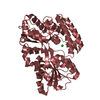
- PDB-7c6t: Crystal structure of beta-glycosides-binding protein (W177X) of A... -
+ Open data

- Basic information
| Entry | Database: PDB / ID: 7c6t | ||||||
|---|---|---|---|---|---|---|---|
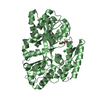
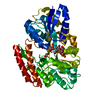
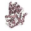
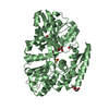
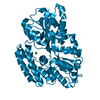
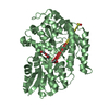
| Title | Crystal structure of beta-glycosides-binding protein (W177X) of ABC transporter in a closed state bound to laminaritriose (Form I) | ||||||
 Components Components | Sugar ABC transporter, periplasmic sugar-binding protein | ||||||
 Keywords Keywords | SUGAR BINDING PROTEIN / Conformational dynamics / substrate-binding protein / Induced-fit mechanism / Two-step ligand binding / Venus Fly-trap mechanism | ||||||
| Function / homology | Bacterial extracellular solute-binding protein / maltose binding / maltose transport / maltodextrin transmembrane transport / ATP-binding cassette (ABC) transporter complex, substrate-binding subunit-containing / Bacterial extracellular solute-binding protein / CARBON DIOXIDE / Sugar ABC transporter, periplasmic sugar-binding protein Function and homology information Function and homology information | ||||||
| Biological species |   Thermus thermophilus HB8 (bacteria) Thermus thermophilus HB8 (bacteria) | ||||||
| Method |  X-RAY DIFFRACTION / MOLECULAR REPLACEMENT / molecular replacement / Resolution: 2.3 Å X-RAY DIFFRACTION / MOLECULAR REPLACEMENT / molecular replacement / Resolution: 2.3 Å | ||||||
 Authors Authors | Kanaujia, S.P. / Chandravanshi, M. / Samanta, R. | ||||||
| Funding support |  India, 1items India, 1items
| ||||||
 Citation Citation | Journal: J.Mol.Biol. / Year: 2020 Title: Conformational Trapping of a beta-Glucosides-Binding Protein Unveils the Selective Two-Step Ligand-Binding Mechanism of ABC Importers. Authors: Chandravanshi, M. / Samanta, R. / Kanaujia, S.P. | ||||||
| History |
|
- Structure visualization
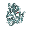
| Structure viewer | Molecule: MolmilJmol/JSmol |
|---|
- Downloads & links
-Download
| PDBx/mmCIF format | 7c6t.cif.gz | 173.9 KB | Display | PDBx/mmCIF format |
|---|---|---|---|---|
| PDB format | pdb7c6t.ent.gz | 135.7 KB | Display | PDB format |
| PDBx/mmJSON format | 7c6t.json.gz | Tree view | PDBx/mmJSON format | |
| Others |  Other downloads Other downloads |
-Validation report
| Arichive directory | https://data.pdbj.org/pub/pdb/validation_reports/c6/7c6tftp://data.pdbj.org/pub/pdb/validation_reports/c6/7c6t | HTTPS FTP |
|---|
-Related structure data
| Related structure data | 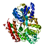 7c63SC  7c64C  7c66C 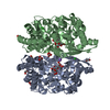 7c67C 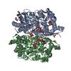 7c68C 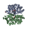 7c69C 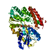 7c6fC 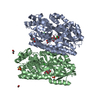 7c6gC  7c6hC 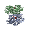 7c6iC 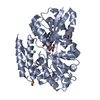 7c6jC 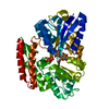 7c6kC 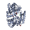 7c6lC 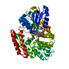 7c6mC 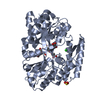 7c6nC  7c6rC 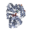 7c6vC 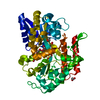 7c6wC  7c6xC 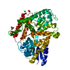 7c6yC  7c6zC 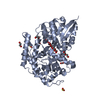 7c70C 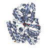 7c71C S: Starting model for refinement C: citing same article ( |
|---|---|
| Similar structure data |


-Links
 PDBj
PDBj


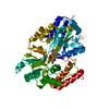
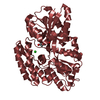
- Assembly
| Deposited unit | 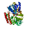
| ||||||||
|---|---|---|---|---|---|---|---|---|---|
| 1 |
| ||||||||
| Unit cell |
|
-Components
-Protein / Sugars , 2 types, 2 molecules A
| #1: Protein | Mass: 45906.211 Da / Num. of mol.: 1 / Mutation: K174R, N175T, S176P, W177del, D178R, V179T Source method: isolated from a genetically manipulated source Source: (gene. exp.) Thermus thermophilus HB8 (bacteria) / Strain: HB8 / Gene: TTHB082 / Plasmid: pET22b / Production host: |
|---|---|
| #2: Polysaccharide | beta-D-glucopyranose-(1-3)-beta-D-glucopyranose-(1-3)-beta-D-glucopyranose Source method: isolated from a genetically manipulated source |
-Non-polymers , 5 types, 145 molecules 


| #3: Chemical | ChemComp-CL /  Mass: 35.453 Da / Num. of mol.: 1 / Source method: obtained synthetically / Formula: Cl Mass: 35.453 Da / Num. of mol.: 1 / Source method: obtained synthetically / Formula: Cl | ||||||
|---|---|---|---|---|---|---|---|
| #4: Chemical |  Mass: 44.010 Da / Num. of mol.: 2 / Source method: obtained synthetically / Formula: CO2 Mass: 44.010 Da / Num. of mol.: 2 / Source method: obtained synthetically / Formula: CO2#5: Chemical | ChemComp-SO4 / |  Mass: 96.063 Da / Num. of mol.: 1 / Source method: obtained synthetically / Formula: SO4 Mass: 96.063 Da / Num. of mol.: 1 / Source method: obtained synthetically / Formula: SO4#6: Chemical |  Mass: 62.068 Da / Num. of mol.: 2 / Source method: obtained synthetically / Formula: C2H6O2 Mass: 62.068 Da / Num. of mol.: 2 / Source method: obtained synthetically / Formula: C2H6O2#7: Water | ChemComp-HOH / | Mass: 18.015 Da / Num. of mol.: 139 / Source method: isolated from a natural source / Formula: H2O |
-Details
| Has ligand of interest | Y |
|---|---|
| Has protein modification | Y |
-Experimental details
-Experiment
| Experiment | Method: X-RAY DIFFRACTION / Number of used crystals: 1 |
|---|
- Sample preparation
| Crystal | Density Matthews: 2.21 Å3/Da / Density % sol: 44.46 % / Description: Orthorhombic |
|---|---|
| Crystal grow | Temperature: 293 K / Method: microbatch / Details: 0.2 M Ammonium sulphate, 40% PEG 8000 |
-Data collection
| Diffraction | Mean temperature: 100 K / Serial crystal experiment: N | ||||||||||||||||||||||||||||||
|---|---|---|---|---|---|---|---|---|---|---|---|---|---|---|---|---|---|---|---|---|---|---|---|---|---|---|---|---|---|---|---|
| Diffraction source | Source: ROTATING ANODE / Type: RIGAKU MICROMAX-007 HF / Wavelength: 1.5418 Å | ||||||||||||||||||||||||||||||
| Detector | Type: RIGAKU RAXIS IV++ / Detector: IMAGE PLATE / Date: Sep 18, 2019 / Details: VariMax HF | ||||||||||||||||||||||||||||||
| Radiation | Protocol: SINGLE WAVELENGTH / Monochromatic (M) / Laue (L): M / Scattering type: x-ray | ||||||||||||||||||||||||||||||
| Radiation wavelength | Wavelength: 1.5418 Å / Relative weight: 1 | ||||||||||||||||||||||||||||||
| Reflection | Resolution: 2.3→57.93 Å / Num. obs: 18713 / % possible obs: 99.8 % / Redundancy: 8.8 % / CC1/2: 0.996 / Rmerge(I) obs: 0.105 / Rpim(I) all: 0.037 / Rrim(I) all: 0.112 / Net I/σ(I): 16.1 / Num. measured all: 165548 / Scaling rejects: 684 | ||||||||||||||||||||||||||||||
| Reflection shell | Diffraction-ID: 1
|
-Phasing
| Phasing | Method: molecular replacement | ||||||
|---|---|---|---|---|---|---|---|
| Phasing MR | Model details: Phaser MODE: MR_AUTO
|
- Processing
| Software |
| ||||||||||||||||||||||||||||||||||||||||||||||||||||||||||||
|---|---|---|---|---|---|---|---|---|---|---|---|---|---|---|---|---|---|---|---|---|---|---|---|---|---|---|---|---|---|---|---|---|---|---|---|---|---|---|---|---|---|---|---|---|---|---|---|---|---|---|---|---|---|---|---|---|---|---|---|---|---|
| Refinement | Method to determine structure: MOLECULAR REPLACEMENT Starting model: 7C63 Resolution: 2.3→55.06 Å / Cor.coef. Fo:Fc: 0.921 / Cor.coef. Fo:Fc free: 0.89 / SU B: 11.674 / SU ML: 0.151 / SU R Cruickshank DPI: 0.0882 / Cross valid method: THROUGHOUT / σ(F): 0 / ESU R: 0.088 / ESU R Free: 0.057 / Stereochemistry target values: MAXIMUM LIKELIHOOD Details: U VALUES : WITH TLS ADDED HYDROGENS HAVE BEEN ADDED IN THE RIDING POSITIONS
| ||||||||||||||||||||||||||||||||||||||||||||||||||||||||||||
| Solvent computation | Ion probe radii: 0.8 Å / Shrinkage radii: 0.8 Å / VDW probe radii: 1.2 Å / Solvent model: MASK | ||||||||||||||||||||||||||||||||||||||||||||||||||||||||||||
| Displacement parameters | Biso max: 70.22 Å2 / Biso mean: 24.888 Å2 / Biso min: 9.34 Å2
| ||||||||||||||||||||||||||||||||||||||||||||||||||||||||||||
| Refinement step | Cycle: final / Resolution: 2.3→55.06 Å
| ||||||||||||||||||||||||||||||||||||||||||||||||||||||||||||
| Refine LS restraints |
| ||||||||||||||||||||||||||||||||||||||||||||||||||||||||||||
| LS refinement shell | Resolution: 2.3→2.36 Å / Rfactor Rfree error: 0 / Total num. of bins used: 20
| ||||||||||||||||||||||||||||||||||||||||||||||||||||||||||||
| Refinement TLS params. | Method: refined / Origin x: 21.0734 Å / Origin y: 1.0922 Å / Origin z: 16.1819 Å
|
