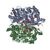
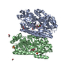
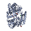
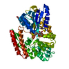
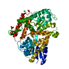
PDB-7c63: Crystal structure of beta-glycosides-binding protein of ABC transporter in an open state (Form I) Method: X-RAY DIFFRACTION / Resolution: 1.63 Å
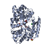
PDB-7c64: Crystal structure of beta-glycosides-binding protein of ABC transporter in an open state (Form II) Method: X-RAY DIFFRACTION / Resolution: 1.63 Å
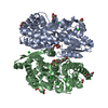
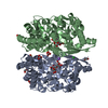
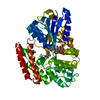
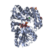
PDB-7c66: Crystal structure of beta-glycosides-binding protein of ABC transporter in a closed state bound to cellobiose Method: X-RAY DIFFRACTION / Resolution: 2.3 Å
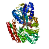
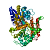
PDB-7c67: Crystal structure of beta-glycosides-binding protein of ABC transporter in a closed state bound to cellotriose Method: X-RAY DIFFRACTION / Resolution: 2 Å
PDB-7c68: Crystal structure of beta-glycosides-binding protein of ABC transporter in a closed state bound to cellotetraose Method: X-RAY DIFFRACTION / Resolution: 2.05 Å
PDB-7c69: Crystal structure of beta-glycosides-binding protein of ABC transporter in a closed state bound to sophorose Method: X-RAY DIFFRACTION / Resolution: 2.35 Å
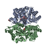
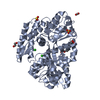
PDB-7c6f: Crystal structure of beta-glycosides-binding protein (W177X) of ABC transporter in an open state Method: X-RAY DIFFRACTION / Resolution: 1.7 Å
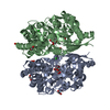
PDB-7c6g: Crystal structure of beta-glycosides-binding protein (W177X) of ABC transporter in an open-liganded state bound to gentiobiose Method: X-RAY DIFFRACTION / Resolution: 1.9 Å
PDB-7c6h: Crystal structure of beta-glycosides-binding protein (W177X) of ABC transporter in an open-liganded state bound to laminaribiose Method: X-RAY DIFFRACTION / Resolution: 1.85 Å
PDB-7c6i: Crystal structure of beta-glycosides-binding protein (W177X) of ABC transporter in an open-liganded state bound to sophorose Method: X-RAY DIFFRACTION / Resolution: 1.7 Å
PDB-7c6j: Crystal structure of beta-glycosides-binding protein (W177X) of ABC transporter in a closed state bound to cellobiose Method: X-RAY DIFFRACTION / Resolution: 2.1 Å
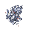
PDB-7c6k: Crystal structure of beta-glycosides-binding protein (W177X) of ABC transporter in a closed state bound to cellotriose (Form I) Method: X-RAY DIFFRACTION / Resolution: 1.77 Å
PDB-7c6l: Crystal structure of beta-glycosides-binding protein (W177X) of ABC transporter in a closed state bound to cellotriose (Form II) Method: X-RAY DIFFRACTION / Resolution: 2.4 Å
PDB-7c6m: Crystal structure of beta-glycosides-binding protein (W177X) of ABC transporter in a closed state bound to cellotetraose (Form I) Method: X-RAY DIFFRACTION / Resolution: 1.9 Å
PDB-7c6n: Crystal structure of beta-glycosides-binding protein (W177X) of ABC transporter in a closed state bound to cellotetraose (Form II) Method: X-RAY DIFFRACTION / Resolution: 2.05 Å
PDB-7c6r: Crystal structure of beta-glycosides-binding protein (W177X) of ABC transporter in a closed state bound to cellopentaose Method: X-RAY DIFFRACTION / Resolution: 1.96 Å
PDB-7c6t: Crystal structure of beta-glycosides-binding protein (W177X) of ABC transporter in a closed state bound to laminaritriose (Form I) Method: X-RAY DIFFRACTION / Resolution: 2.3 Å
PDB-7c6v: Crystal structure of beta-glycosides-binding protein (W177X) of ABC transporter in a closed state bound to laminaritriose (Form II) Method: X-RAY DIFFRACTION / Resolution: 2.8 Å
PDB-7c6w: Crystal structure of beta-glycosides-binding protein (W177X) of ABC transporter in a closed state bound to laminaritetraose Method: X-RAY DIFFRACTION / Resolution: 1.88 Å
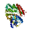
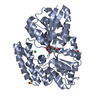
PDB-7c6x: Crystal structure of beta-glycosides-binding protein (W41A) of ABC transporter in an open state (Form I) Method: X-RAY DIFFRACTION / Resolution: 2.65 Å
PDB-7c6y: Crystal structure of beta-glycosides-binding protein (W41A) of ABC transporter in an open state (Form II) Method: X-RAY DIFFRACTION / Resolution: 1.7 Å
PDB-7c6z: Crystal structure of beta-glycosides-binding protein (W67A) of ABC transporter in an open state Method: X-RAY DIFFRACTION / Resolution: 1.63 Å
PDB-7c70: Crystal structure of beta-glycosides-binding protein (W67A) of ABC transporter in an open-liganded state bound to gentiobiose Method: X-RAY DIFFRACTION / Resolution: 1.63 Å
PDB-7c71: Crystal structure of beta-glycosides-binding protein (E117A) of ABC transporter in an open state Method: X-RAY DIFFRACTION / Resolution: 2.1 Å
In the structure databanks used in Yorodumi, some data are registered as the other names, "COVID-19 virus" and "2019-nCoV". Here are the details of the virus and the list of structure data.
Jan 31, 2019. EMDB accession codes are about to change! (news from PDBe EMDB page)
EMDB accession codes are about to change! (news from PDBe EMDB page)
The allocation of 4 digits for EMDB accession codes will soon come to an end. Whilst these codes will remain in use, new EMDB accession codes will include an additional digit and will expand incrementally as the available range of codes is exhausted. The current 4-digit format prefixed with “EMD-” (i.e. EMD-XXXX) will advance to a 5-digit format (i.e. EMD-XXXXX), and so on. It is currently estimated that the 4-digit codes will be depleted around Spring 2019, at which point the 5-digit format will come into force.
The EM Navigator/Yorodumi systems omit the EMD- prefix.
Related info.:Q: What is EMD? / ID/Accession-code notation in Yorodumi/EM Navigator
 Movie
Movie Controller
 Structure viewers
 About Yorodumi Papers


 Authors
 External links
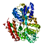


 Keywords
 thermus thermophilus (strain hb8 / atcc 27634 / dsm 579) (bacteria)