 Movie
Movie Controller

[English] 日本語
 Yorodumi
- PDB-6zh7: Crystal structure of fatty acid photodecarboxylase in the dark st... -
+ Open data

- Basic information
| Entry | Database: PDB / ID: 6zh7 | |||||||||
|---|---|---|---|---|---|---|---|---|---|---|
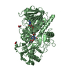
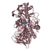
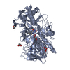
| Title | Crystal structure of fatty acid photodecarboxylase in the dark state determined by serial femtosecond crystallography at room temperature | |||||||||
 Components Components | Fatty acid photodecarboxylase, chloroplastic | |||||||||
 Keywords Keywords | OXIDOREDUCTASE / photoenzyme / XFEL / radiation damage free | |||||||||
| Function / homology |  Function and homology information Function and homology informationfatty acid photodecarboxylase / choline dehydrogenase activity / : / chloroplast / flavin adenine dinucleotide binding / lyase activity / membrane Similarity search - Function | |||||||||
| Biological species |  Chlorella variabilis (plant) Chlorella variabilis (plant) | |||||||||
| Method |  X-RAY DIFFRACTION / FREE ELECTRON LASER / MOLECULAR REPLACEMENT / Resolution: 2 Å X-RAY DIFFRACTION / FREE ELECTRON LASER / MOLECULAR REPLACEMENT / Resolution: 2 Å | |||||||||
 Authors Authors | Hadjidemetriou, K. / Coquelle, N. / Weik, M. / Schlichting, I. / Barends, T.R.M. / Colletier, J.P. | |||||||||
| Funding support |  France, 2items France, 2items
| |||||||||
 Citation Citation | Journal: Science / Year: 2021 Title: Mechanism and dynamics of fatty acid photodecarboxylase. Authors: Sorigue, D. / Hadjidemetriou, K. / Blangy, S. / Gotthard, G. / Bonvalet, A. / Coquelle, N. / Samire, P. / Aleksandrov, A. / Antonucci, L. / Benachir, A. / Boutet, S. / Byrdin, M. / ...Authors: Sorigue, D. / Hadjidemetriou, K. / Blangy, S. / Gotthard, G. / Bonvalet, A. / Coquelle, N. / Samire, P. / Aleksandrov, A. / Antonucci, L. / Benachir, A. / Boutet, S. / Byrdin, M. / Cammarata, M. / Carbajo, S. / Cuine, S. / Doak, R.B. / Foucar, L. / Gorel, A. / Grunbein, M. / Hartmann, E. / Hienerwadel, R. / Hilpert, M. / Kloos, M. / Lane, T.J. / Legeret, B. / Legrand, P. / Li-Beisson, Y. / Moulin, S.L.Y. / Nurizzo, D. / Peltier, G. / Schiro, G. / Shoeman, R.L. / Sliwa, M. / Solinas, X. / Zhuang, B. / Barends, T.R.M. / Colletier, J.P. / Joffre, M. / Royant, A. / Berthomieu, C. / Weik, M. / Domratcheva, T. / Brettel, K. / Vos, M.H. / Schlichting, I. / Arnoux, P. / Muller, P. / Beisson, F. #1: Journal: Science / Year: 2021Title: Mechanism and dynamics of fatty acid photodecarboxylase Authors: Sorigue, D. / Hadjidemetriou, K. / Blangy, S. / Gotthard, G. / Bonvalet, A. / Coquelle, N. / Samire, P. / Aleksandrov, A. / Antonucci, L. / Benachir, A. / Boutet, S. / Byrdin, M. / ...Authors: Sorigue, D. / Hadjidemetriou, K. / Blangy, S. / Gotthard, G. / Bonvalet, A. / Coquelle, N. / Samire, P. / Aleksandrov, A. / Antonucci, L. / Benachir, A. / Boutet, S. / Byrdin, M. / Cammarata, M. / Carbajo, S. / Cuine, S. / Doak, R.B. / Foucar, L. / Gorel, A. / Grunbein, M. / Hartmann, E. / Hienerwadel, R. / Hilpert, M. / Kloos, M. / Lane, T.J. / Legeret, B. / Legrand, P. / Li-Beisson, Y. / Moulin, S. / Nurizzo, D. / Peltier, G. / Schiro, G. / Shoeman, R.L. / Sliwa, M. / Solinas, X. / Zhuang, B. / Barends, T.R.M. / Colletier, J.P. / Joffre, M. / Royant, A. / Berthomieu, C. / Weik, M. / Domratcheva, T. / Brettel, K. / Vos, M.H. / Schlichting, I. / Arnoux, P. / Muller, P. / Beisson, F. | |||||||||
| History |
|
- Structure visualization
| Structure viewer | Molecule: MolmilJmol/JSmol |
|---|
- Downloads & links
-Download
| PDBx/mmCIF format | 6zh7.cif.gz | 242.4 KB | Display | PDBx/mmCIF format |
|---|---|---|---|---|
| PDB format | pdb6zh7.ent.gz | 189.7 KB | Display | PDB format |
| PDBx/mmJSON format | 6zh7.json.gz | Tree view | PDBx/mmJSON format | |
| Others |  Other downloads Other downloads |
-Validation report
| Arichive directory | https://data.pdbj.org/pub/pdb/validation_reports/zh/6zh7ftp://data.pdbj.org/pub/pdb/validation_reports/zh/6zh7 | HTTPS FTP |
|---|
-Related structure data
| Related structure data |  6yruSC 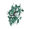 6yrvC 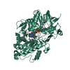 6yrxC 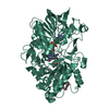 6yrzC  6ys1C 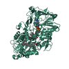 6ys2C 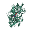 7av4C S: Starting model for refinement C: citing same article ( |
|---|---|
| Similar structure data | |
| Experimental dataset #1 | Data reference: 10.11577/1775333 / Data set type: diffraction image data |


-Links
 PDBj
PDBj

- Assembly
| Deposited unit | 
| ||||||||||||||||||
|---|---|---|---|---|---|---|---|---|---|---|---|---|---|---|---|---|---|---|---|
| 1 | 
| ||||||||||||||||||
| 2 | 
| ||||||||||||||||||
| Unit cell |
| ||||||||||||||||||
| Noncrystallographic symmetry (NCS) | NCS domain:
NCS domain segments: Component-ID: _ / Ens-ID: 1 / Beg auth comp-ID: SER / Beg label comp-ID: SER / End auth comp-ID: GLY / End label comp-ID: GLY / Refine code: _ / Auth seq-ID: 77 - 643 / Label seq-ID: 1 - 567
|
-Components
| #1: Protein | Mass: 61116.598 Da / Num. of mol.: 2 Source method: isolated from a genetically manipulated source Source: (gene. exp.) Chlorella variabilis (plant) / Gene: FAP, CHLNCDRAFT_28598 / Production host:  References: UniProt: A0A248QE08, fatty acid photodecarboxylase #2: Chemical |   Mass: 785.550 Da / Num. of mol.: 2 / Source method: obtained synthetically / Formula: C27H33N9O15P2 / Feature type: SUBJECT OF INVESTIGATION / Comment: FAD*YM Mass: 785.550 Da / Num. of mol.: 2 / Source method: obtained synthetically / Formula: C27H33N9O15P2 / Feature type: SUBJECT OF INVESTIGATION / Comment: FAD*YM#3: Chemical | ChemComp-STE /   Mass: 284.477 Da / Num. of mol.: 4 / Source method: obtained synthetically / Formula: C18H36O2 / Feature type: SUBJECT OF INVESTIGATION Mass: 284.477 Da / Num. of mol.: 4 / Source method: obtained synthetically / Formula: C18H36O2 / Feature type: SUBJECT OF INVESTIGATION#4: Water | ChemComp-HOH / |  Mass: 18.015 Da / Num. of mol.: 394 / Source method: isolated from a natural source / Formula: H2O Mass: 18.015 Da / Num. of mol.: 394 / Source method: isolated from a natural source / Formula: H2OHas ligand of interest | Y | |
|---|
-Experimental details
-Experiment
| Experiment | Method: X-RAY DIFFRACTION / Number of used crystals: 1 |
|---|
- Sample preparation
| Crystal | Density Matthews: 2.76 Å3/Da / Density % sol: 55.53 % |
|---|---|
| Crystal grow | Temperature: 281.15 K / Method: batch mode / pH: 5.5 Details: 19% (w/v) PEG 4000, 0.1 M sodium citrate pH 5.5, 10 mM spermidine |
-Data collection
| Diffraction | Mean temperature: 277.15 K / Serial crystal experiment: Y |
|---|---|
| Diffraction source | Source: FREE ELECTRON LASER / Site: SLAC LCLS  / Beamline: CXI / Wavelength: 1.3 Å / Beamline: CXI / Wavelength: 1.3 Å |
| Detector | Type: CS-PAD CXI-1 / Detector: PIXEL / Date: Nov 22, 2018 |
| Radiation | Protocol: SINGLE WAVELENGTH / Monochromatic (M) / Laue (L): M / Scattering type: x-ray |
| Radiation wavelength | Wavelength: 1.3 Å / Relative weight: 1 |
| Reflection | Resolution: 2→25 Å / Num. obs: 93061 / % possible obs: 100 % / Redundancy: 355 % / CC1/2: 0.982 / CC star: 0.996 / R split: 0.151 / Net I/σ(I): 5.6 |
| Reflection shell | Resolution: 2→2.05 Å / Redundancy: 236 % / Num. unique obs: 6086 / CC1/2: 0.548 / CC star: 0.841 / R split: 0.685 / % possible all: 100 |
| Serial crystallography sample delivery | Method: injection |
- Processing
| Software |
| ||||||||||||||||||||||||||||||||||||||||||||||||||||||||||||
|---|---|---|---|---|---|---|---|---|---|---|---|---|---|---|---|---|---|---|---|---|---|---|---|---|---|---|---|---|---|---|---|---|---|---|---|---|---|---|---|---|---|---|---|---|---|---|---|---|---|---|---|---|---|---|---|---|---|---|---|---|---|
| Refinement | Method to determine structure: MOLECULAR REPLACEMENT Starting model: 6YRU Resolution: 2→24.95 Å / Cor.coef. Fo:Fc: 0.951 / Cor.coef. Fo:Fc free: 0.932 / SU B: 5.893 / SU ML: 0.151 / Cross valid method: FREE R-VALUE / σ(F): 0 / ESU R: 0.173 / ESU R Free: 0.154 / Stereochemistry target values: MAXIMUM LIKELIHOOD Details: HYDROGENS HAVE BEEN ADDED IN THE RIDING POSITIONS U VALUES : REFINED INDIVIDUALLY
| ||||||||||||||||||||||||||||||||||||||||||||||||||||||||||||
| Solvent computation | Ion probe radii: 0.8 Å / Shrinkage radii: 0.8 Å / VDW probe radii: 1.2 Å / Solvent model: MASK | ||||||||||||||||||||||||||||||||||||||||||||||||||||||||||||
| Displacement parameters | Biso max: 103.15 Å2 / Biso mean: 29.803 Å2 / Biso min: 11.55 Å2
| ||||||||||||||||||||||||||||||||||||||||||||||||||||||||||||
| Refinement step | Cycle: final / Resolution: 2→24.95 Å
| ||||||||||||||||||||||||||||||||||||||||||||||||||||||||||||
| Refine LS restraints |
| ||||||||||||||||||||||||||||||||||||||||||||||||||||||||||||
| Refine LS restraints NCS | Ens-ID: 1 / Number: 18263 / Refine-ID: X-RAY DIFFRACTION / Type: interatomic distance / Rms dev position: 0.04 Å / Weight position: 0.05
| ||||||||||||||||||||||||||||||||||||||||||||||||||||||||||||
| LS refinement shell | Resolution: 2→2.052 Å / Rfactor Rfree error: 0 / Total num. of bins used: 20
|
