 Movie
Movie Controller


+ Open data

- Basic information
| Entry | Database: PDB / ID: 6yrv | ||||||
|---|---|---|---|---|---|---|---|
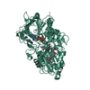
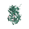
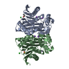
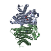
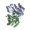
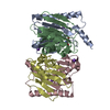
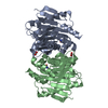
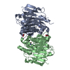
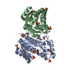
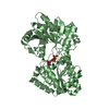
| Title | Crystal structure of FAP after illumination at 100K | ||||||
 Components Components | Fatty acid Photodecarboxylase | ||||||
 Keywords Keywords | BIOSYNTHETIC PROTEIN / GMC fold | ||||||
| Function / homology |  Function and homology information Function and homology informationfatty acid photodecarboxylase / choline dehydrogenase activity / : / chloroplast / flavin adenine dinucleotide binding / lyase activity / membrane Similarity search - Function | ||||||
| Biological species |  Chlorella variabilis (plant) Chlorella variabilis (plant) | ||||||
| Method |  X-RAY DIFFRACTION / SYNCHROTRON / MOLECULAR REPLACEMENT / Resolution: 1.94 Å X-RAY DIFFRACTION / SYNCHROTRON / MOLECULAR REPLACEMENT / Resolution: 1.94 Å | ||||||
 Authors Authors | Sorigue, D. / Gotthard, G. / Blangy, S. / Nurizzo, D. / Royant, A. / Beisson, F. / Arnoux, P. | ||||||
| Funding support |  France, 1items France, 1items
| ||||||
 Citation Citation | Journal: Science / Year: 2021 Title: Mechanism and dynamics of fatty acid photodecarboxylase. Authors: Sorigue, D. / Hadjidemetriou, K. / Blangy, S. / Gotthard, G. / Bonvalet, A. / Coquelle, N. / Samire, P. / Aleksandrov, A. / Antonucci, L. / Benachir, A. / Boutet, S. / Byrdin, M. / ...Authors: Sorigue, D. / Hadjidemetriou, K. / Blangy, S. / Gotthard, G. / Bonvalet, A. / Coquelle, N. / Samire, P. / Aleksandrov, A. / Antonucci, L. / Benachir, A. / Boutet, S. / Byrdin, M. / Cammarata, M. / Carbajo, S. / Cuine, S. / Doak, R.B. / Foucar, L. / Gorel, A. / Grunbein, M. / Hartmann, E. / Hienerwadel, R. / Hilpert, M. / Kloos, M. / Lane, T.J. / Legeret, B. / Legrand, P. / Li-Beisson, Y. / Moulin, S.L.Y. / Nurizzo, D. / Peltier, G. / Schiro, G. / Shoeman, R.L. / Sliwa, M. / Solinas, X. / Zhuang, B. / Barends, T.R.M. / Colletier, J.P. / Joffre, M. / Royant, A. / Berthomieu, C. / Weik, M. / Domratcheva, T. / Brettel, K. / Vos, M.H. / Schlichting, I. / Arnoux, P. / Muller, P. / Beisson, F. | ||||||
| History |
|
- Structure visualization
| Structure viewer | Molecule: MolmilJmol/JSmol |
|---|
- Downloads & links
-Download
| PDBx/mmCIF format | 6yrv.cif.gz | 143.9 KB | Display | PDBx/mmCIF format |
|---|---|---|---|---|
| PDB format | pdb6yrv.ent.gz | Display | PDB format | |
| PDBx/mmJSON format | 6yrv.json.gz | Tree view | PDBx/mmJSON format | |
| Others |  Other downloads Other downloads |
-Validation report
| Arichive directory | https://data.pdbj.org/pub/pdb/validation_reports/yr/6yrvftp://data.pdbj.org/pub/pdb/validation_reports/yr/6yrv | HTTPS FTP |
|---|
-Related structure data
| Related structure data |  6yruSC 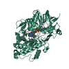 6yrxC 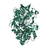 6yrzC  6ys1C 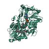 6ys2C  6zh7C 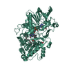 7av4C S: Starting model for refinement C: citing same article ( |
|---|---|
| Similar structure data | |
| Experimental dataset #1 | Data reference: 10.11577/1775333 / Data set type: diffraction image data |
-Links
 PDBj
PDBj


- Assembly
| Deposited unit | 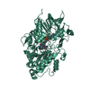
| ||||||||
|---|---|---|---|---|---|---|---|---|---|
| 1 |
| ||||||||
| Unit cell |
|
-Components
-Protein , 1 types, 1 molecules AAA
| #1: Protein | Mass: 61086.574 Da / Num. of mol.: 1 Source method: isolated from a genetically manipulated source Source: (gene. exp.) Chlorella variabilis (plant) / Production host:  |
|---|
-Non-polymers , 5 types, 528 molecules 


| #2: Chemical | ChemComp-FAD /  Mass: 785.550 Da / Num. of mol.: 1 / Source method: obtained synthetically / Formula: C27H33N9O15P2 / Comment: FAD*YM Mass: 785.550 Da / Num. of mol.: 1 / Source method: obtained synthetically / Formula: C27H33N9O15P2 / Comment: FAD*YM |
|---|---|
| #3: Chemical | ChemComp-PJ8 /  Mass: 240.468 Da / Num. of mol.: 1 / Source method: obtained synthetically / Formula: C17H36 / Feature type: SUBJECT OF INVESTIGATION Mass: 240.468 Da / Num. of mol.: 1 / Source method: obtained synthetically / Formula: C17H36 / Feature type: SUBJECT OF INVESTIGATION |
| #4: Chemical | ChemComp-CO2 /  Mass: 44.010 Da / Num. of mol.: 1 / Source method: obtained synthetically / Formula: CO2 Mass: 44.010 Da / Num. of mol.: 1 / Source method: obtained synthetically / Formula: CO2 |
| #5: Chemical | ChemComp-STE /  Mass: 284.477 Da / Num. of mol.: 1 / Source method: obtained synthetically / Formula: C18H36O2 Mass: 284.477 Da / Num. of mol.: 1 / Source method: obtained synthetically / Formula: C18H36O2 |
| #6: Water | ChemComp-HOH / Mass: 18.015 Da / Num. of mol.: 524 / Source method: isolated from a natural source / Formula: H2O |
-Details
| Has ligand of interest | Y |
|---|
-Experimental details
-Experiment
| Experiment | Method: X-RAY DIFFRACTION / Number of used crystals: 1 |
|---|
- Sample preparation
| Crystal | Density Matthews: 3.01 Å3/Da / Density % sol: 59.12 % |
|---|---|
| Crystal grow | Temperature: 281 K / Method: vapor diffusion, sitting drop / pH: 5.5 Details: PEG 4000 25-40%, Na citrate 100 mM, spermidine 10mM |
-Data collection
| Diffraction | Mean temperature: 100 K / Serial crystal experiment: N | ||||||||||||||||||||||||||||||||||||||||||||||||||||||||||||||||||||||||||||||||||||||||||||||||||||||||||||||||||||||||||||||||||||||||||||||||||||||||||||||||||||||||
|---|---|---|---|---|---|---|---|---|---|---|---|---|---|---|---|---|---|---|---|---|---|---|---|---|---|---|---|---|---|---|---|---|---|---|---|---|---|---|---|---|---|---|---|---|---|---|---|---|---|---|---|---|---|---|---|---|---|---|---|---|---|---|---|---|---|---|---|---|---|---|---|---|---|---|---|---|---|---|---|---|---|---|---|---|---|---|---|---|---|---|---|---|---|---|---|---|---|---|---|---|---|---|---|---|---|---|---|---|---|---|---|---|---|---|---|---|---|---|---|---|---|---|---|---|---|---|---|---|---|---|---|---|---|---|---|---|---|---|---|---|---|---|---|---|---|---|---|---|---|---|---|---|---|---|---|---|---|---|---|---|---|---|---|---|---|---|---|---|---|
| Diffraction source | Source: SYNCHROTRON / Site: ESRF / Beamline: ID29 / Wavelength: 0.97372 Å | ||||||||||||||||||||||||||||||||||||||||||||||||||||||||||||||||||||||||||||||||||||||||||||||||||||||||||||||||||||||||||||||||||||||||||||||||||||||||||||||||||||||||
| Detector | Type: DECTRIS EIGER X 4M / Detector: PIXEL / Date: Mar 10, 2018 | ||||||||||||||||||||||||||||||||||||||||||||||||||||||||||||||||||||||||||||||||||||||||||||||||||||||||||||||||||||||||||||||||||||||||||||||||||||||||||||||||||||||||
| Radiation | Protocol: SINGLE WAVELENGTH / Monochromatic (M) / Laue (L): M / Scattering type: x-ray | ||||||||||||||||||||||||||||||||||||||||||||||||||||||||||||||||||||||||||||||||||||||||||||||||||||||||||||||||||||||||||||||||||||||||||||||||||||||||||||||||||||||||
| Radiation wavelength | Wavelength: 0.97372 Å / Relative weight: 1 | ||||||||||||||||||||||||||||||||||||||||||||||||||||||||||||||||||||||||||||||||||||||||||||||||||||||||||||||||||||||||||||||||||||||||||||||||||||||||||||||||||||||||
| Reflection | Resolution: 1.94→86.5 Å / Num. obs: 54600 / % possible obs: 100 % / Redundancy: 6.585 % / Biso Wilson estimate: 34.131 Å2 / CC1/2: 0.998 / Rmerge(I) obs: 0.106 / Rrim(I) all: 0.116 / Χ2: 1.02 / Net I/σ(I): 11.62 | ||||||||||||||||||||||||||||||||||||||||||||||||||||||||||||||||||||||||||||||||||||||||||||||||||||||||||||||||||||||||||||||||||||||||||||||||||||||||||||||||||||||||
| Reflection shell | Diffraction-ID: 1
|
- Processing
| Software |
| |||||||||||||||||||||||||||||||||||||||||||||||||||||||||||||||||||||||||||||||||||||||||||||||||||||||||||||||||||||||||||||||||||||||||||||||||||||||||||||||||||||||||||||||||||||||||||||||||||||||||||||||||||||||||||||||||||||||
|---|---|---|---|---|---|---|---|---|---|---|---|---|---|---|---|---|---|---|---|---|---|---|---|---|---|---|---|---|---|---|---|---|---|---|---|---|---|---|---|---|---|---|---|---|---|---|---|---|---|---|---|---|---|---|---|---|---|---|---|---|---|---|---|---|---|---|---|---|---|---|---|---|---|---|---|---|---|---|---|---|---|---|---|---|---|---|---|---|---|---|---|---|---|---|---|---|---|---|---|---|---|---|---|---|---|---|---|---|---|---|---|---|---|---|---|---|---|---|---|---|---|---|---|---|---|---|---|---|---|---|---|---|---|---|---|---|---|---|---|---|---|---|---|---|---|---|---|---|---|---|---|---|---|---|---|---|---|---|---|---|---|---|---|---|---|---|---|---|---|---|---|---|---|---|---|---|---|---|---|---|---|---|---|---|---|---|---|---|---|---|---|---|---|---|---|---|---|---|---|---|---|---|---|---|---|---|---|---|---|---|---|---|---|---|---|---|---|---|---|---|---|---|---|---|---|---|---|---|---|---|---|---|
| Refinement | Method to determine structure: MOLECULAR REPLACEMENT Starting model: 6YRU Resolution: 1.94→86 Å / Cor.coef. Fo:Fc: 0.968 / Cor.coef. Fo:Fc free: 0.955 / SU B: 3.572 / SU ML: 0.098 / Cross valid method: FREE R-VALUE / ESU R: 0.127 / ESU R Free: 0.124 Details: Hydrogens have been added in their riding positions
| |||||||||||||||||||||||||||||||||||||||||||||||||||||||||||||||||||||||||||||||||||||||||||||||||||||||||||||||||||||||||||||||||||||||||||||||||||||||||||||||||||||||||||||||||||||||||||||||||||||||||||||||||||||||||||||||||||||||
| Solvent computation | Ion probe radii: 0.8 Å / Shrinkage radii: 0.8 Å / VDW probe radii: 1.2 Å | |||||||||||||||||||||||||||||||||||||||||||||||||||||||||||||||||||||||||||||||||||||||||||||||||||||||||||||||||||||||||||||||||||||||||||||||||||||||||||||||||||||||||||||||||||||||||||||||||||||||||||||||||||||||||||||||||||||||
| Displacement parameters | Biso mean: 29.774 Å2
| |||||||||||||||||||||||||||||||||||||||||||||||||||||||||||||||||||||||||||||||||||||||||||||||||||||||||||||||||||||||||||||||||||||||||||||||||||||||||||||||||||||||||||||||||||||||||||||||||||||||||||||||||||||||||||||||||||||||
| Refinement step | Cycle: LAST / Resolution: 1.94→86 Å
| |||||||||||||||||||||||||||||||||||||||||||||||||||||||||||||||||||||||||||||||||||||||||||||||||||||||||||||||||||||||||||||||||||||||||||||||||||||||||||||||||||||||||||||||||||||||||||||||||||||||||||||||||||||||||||||||||||||||
| Refine LS restraints |
| |||||||||||||||||||||||||||||||||||||||||||||||||||||||||||||||||||||||||||||||||||||||||||||||||||||||||||||||||||||||||||||||||||||||||||||||||||||||||||||||||||||||||||||||||||||||||||||||||||||||||||||||||||||||||||||||||||||||
| LS refinement shell | Refine-ID: X-RAY DIFFRACTION / Total num. of bins used: 20
|
