 Movie
Movie Controller


+ Open data

- Basic information
| Entry | Database: PDB / ID: 6zd5 | |||||||||
|---|---|---|---|---|---|---|---|---|---|---|
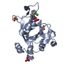
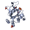
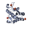
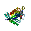
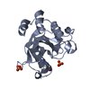
| Title | Crystal structure of YTHDC1 S378A mutant complex with m6A | |||||||||
 Components Components | YTH domain containing 1 | |||||||||
 Keywords Keywords | RNA BINDING PROTEIN / YTHDC1 / m6A / complex / inhibitor | |||||||||
| Function / homology | N-methyladenosine Function and homology information Function and homology information | |||||||||
| Biological species |  Homo sapiens (human) Homo sapiens (human) | |||||||||
| Method |  X-RAY DIFFRACTION / SYNCHROTRON / MOLECULAR REPLACEMENT / Resolution: 2.3 Å X-RAY DIFFRACTION / SYNCHROTRON / MOLECULAR REPLACEMENT / Resolution: 2.3 Å | |||||||||
 Authors Authors | Bedi, R.K. / Li, Y. / Caflisch, A. | |||||||||
| Funding support |  Switzerland, Switzerland,  Sweden, 2items Sweden, 2items
| |||||||||
 Citation Citation | Journal: J Chem Theory Comput / Year: 2021 Title: Atomistic and Thermodynamic Analysis of N6-Methyladenosine (m 6 A) Recognition by the Reader Domain of YTHDC1. Authors: Li, Y. / Bedi, R.K. / Wiedmer, L. / Sun, X. / Huang, D. / Caflisch, A. | |||||||||
| History |
|
- Structure visualization
| Structure viewer | Molecule: MolmilJmol/JSmol |
|---|
- Downloads & links
-Download
| PDBx/mmCIF format | 6zd5.cif.gz | 100.9 KB | Display | PDBx/mmCIF format |
|---|---|---|---|---|
| PDB format | pdb6zd5.ent.gz | 61.4 KB | Display | PDB format |
| PDBx/mmJSON format | 6zd5.json.gz | Tree view | PDBx/mmJSON format | |
| Others |  Other downloads Other downloads |
-Validation report
| Arichive directory | https://data.pdbj.org/pub/pdb/validation_reports/zd/6zd5ftp://data.pdbj.org/pub/pdb/validation_reports/zd/6zd5 | HTTPS FTP |
|---|
-Related structure data
| Related structure data | 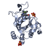 6ynnC  6ynoC  6zcmC  6zcnC 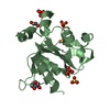 6zd3C  6zd4C  6zd7C  6zd8C  6zd9C  6zdaC  4r3hS S: Starting model for refinement C: citing same article ( |
|---|---|
| Similar structure data |

-Links
 PDBj
PDBj- Assembly
| Deposited unit | 
| ||||||||||||
|---|---|---|---|---|---|---|---|---|---|---|---|---|---|
| 1 | 
| ||||||||||||
| 2 | 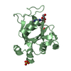
| ||||||||||||
| Unit cell |
|
-Components
| #1: Protein | Mass: 20951.160 Da / Num. of mol.: 2 Source method: isolated from a genetically manipulated source Source: (gene. exp.) Homo sapiens (human) / Gene: YTHDC1 / Production host:  #2: Chemical | ChemComp-SO4 /   Mass: 96.063 Da / Num. of mol.: 7 / Source method: obtained synthetically / Formula: SO4 Mass: 96.063 Da / Num. of mol.: 7 / Source method: obtained synthetically / Formula: SO4#3: Chemical |   Mass: 281.268 Da / Num. of mol.: 2 / Source method: obtained synthetically / Formula: C11H15N5O4 / Feature type: SUBJECT OF INVESTIGATION Mass: 281.268 Da / Num. of mol.: 2 / Source method: obtained synthetically / Formula: C11H15N5O4 / Feature type: SUBJECT OF INVESTIGATION#4: Water | ChemComp-HOH / |  Mass: 18.015 Da / Num. of mol.: 183 / Source method: isolated from a natural source / Formula: H2O Mass: 18.015 Da / Num. of mol.: 183 / Source method: isolated from a natural source / Formula: H2OHas ligand of interest | Y | |
|---|
-Experimental details
-Experiment
| Experiment | Method: X-RAY DIFFRACTION / Number of used crystals: 1 |
|---|
- Sample preparation
| Crystal | Density Matthews: 2.01 Å3/Da / Density % sol: 38.78 % |
|---|---|
| Crystal grow | Temperature: 295 K / Method: vapor diffusion, hanging drop Details: 25% PEG3350, 0.2M Ammonium Sulphate, 0.1M bis-tris, pH 6.5 |
-Data collection
| Diffraction | Mean temperature: 100 K / Serial crystal experiment: N |
|---|---|
| Diffraction source | Source: SYNCHROTRON / Site: SLS / Beamline: X06DA / Wavelength: 1 Å |
| Detector | Type: DECTRIS PILATUS 2M-F / Detector: PIXEL / Date: Jun 17, 2019 |
| Radiation | Protocol: SINGLE WAVELENGTH / Monochromatic (M) / Laue (L): M / Scattering type: x-ray |
| Radiation wavelength | Wavelength: 1 Å / Relative weight: 1 |
| Reflection | Resolution: 2.3→40.89 Å / Num. obs: 14545 / % possible obs: 99.8 % / Redundancy: 4.5 % / Biso Wilson estimate: 22.21 Å2 / CC1/2: 0.998 / Net I/σ(I): 25.77 |
| Reflection shell | Resolution: 2.3→2.44 Å / Num. unique obs: 2296 / CC1/2: 0.993 |
- Processing
| Software |
| ||||||||||||||||||||||||||||||||||||||||||
|---|---|---|---|---|---|---|---|---|---|---|---|---|---|---|---|---|---|---|---|---|---|---|---|---|---|---|---|---|---|---|---|---|---|---|---|---|---|---|---|---|---|---|---|
| Refinement | Method to determine structure: MOLECULAR REPLACEMENT Starting model: 4r3h Resolution: 2.3→40.89 Å / SU ML: 0.2654 / Cross valid method: FREE R-VALUE / σ(F): 1.36 / Phase error: 24.1658 Stereochemistry target values: GeoStd + Monomer Library + CDL v1.2
| ||||||||||||||||||||||||||||||||||||||||||
| Solvent computation | Shrinkage radii: 0.9 Å / VDW probe radii: 1.11 Å / Solvent model: FLAT BULK SOLVENT MODEL | ||||||||||||||||||||||||||||||||||||||||||
| Displacement parameters | Biso mean: 22.87 Å2 | ||||||||||||||||||||||||||||||||||||||||||
| Refinement step | Cycle: LAST / Resolution: 2.3→40.89 Å
| ||||||||||||||||||||||||||||||||||||||||||
| Refine LS restraints |
| ||||||||||||||||||||||||||||||||||||||||||
| LS refinement shell |
|
