 Movie
Movie Controller


+ Open data

- Basic information
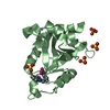
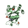
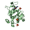
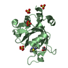
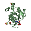
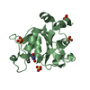
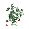
| Entry | Database: PDB / ID: 6we8 | ||||||
|---|---|---|---|---|---|---|---|
| Title | YTH domain of human YTHDC1 | ||||||
 Components Components | YTH domain-containing protein 1 | ||||||
 Keywords Keywords | DNA BINDING PROTEIN / N6-methyladenine binding protein domain | ||||||
| Function / homology |  Function and homology information Function and homology informationmRNA splice site recognition / Nuclear RNA decay / N6-methyladenosine-containing RNA reader activity / dosage compensation by inactivation of X chromosome / regulation of mRNA splicing, via spliceosome / post-transcriptional regulation of gene expression / regulation of alternative mRNA splicing, via spliceosome / brown fat cell differentiation / molecular sequestering activity / mRNA export from nucleus ...mRNA splice site recognition / Nuclear RNA decay / N6-methyladenosine-containing RNA reader activity / dosage compensation by inactivation of X chromosome / regulation of mRNA splicing, via spliceosome / post-transcriptional regulation of gene expression / regulation of alternative mRNA splicing, via spliceosome / brown fat cell differentiation / molecular sequestering activity / mRNA export from nucleus / mRNA splicing, via spliceosome / nuclear speck / mRNA binding / RNA binding / nucleoplasm / nucleus Similarity search - Function | ||||||
| Biological species |  Homo sapiens (human) Homo sapiens (human) | ||||||
| Method |  X-RAY DIFFRACTION / SYNCHROTRON / MOLECULAR REPLACEMENT / Resolution: 1.18 Å X-RAY DIFFRACTION / SYNCHROTRON / MOLECULAR REPLACEMENT / Resolution: 1.18 Å | ||||||
 Authors Authors | Horton, J.R. / Cheng, X. | ||||||
| Funding support |  United States, 1items United States, 1items
| ||||||
 Citation Citation | Journal: Nucleic Acids Res. / Year: 2020 Title: Biochemical and structural basis for YTH domain of human YTHDC1 binding to methylated adenine in DNA. Authors: Woodcock, C.B. / Horton, J.R. / Zhou, J. / Bedford, M.T. / Blumenthal, R.M. / Zhang, X. / Cheng, X. | ||||||
| History |
|
- Structure visualization
| Structure viewer | Molecule: MolmilJmol/JSmol |
|---|
- Downloads & links
-Download
| PDBx/mmCIF format | 6we8.cif.gz | 175.9 KB | Display | PDBx/mmCIF format |
|---|---|---|---|---|
| PDB format | pdb6we8.ent.gz | 121.7 KB | Display | PDB format |
| PDBx/mmJSON format | 6we8.json.gz | Tree view | PDBx/mmJSON format | |
| Others |  Other downloads Other downloads |
-Validation report
| Arichive directory | https://data.pdbj.org/pub/pdb/validation_reports/we/6we8ftp://data.pdbj.org/pub/pdb/validation_reports/we/6we8 | HTTPS FTP |
|---|
-Related structure data
| Related structure data |  6we9C  6weaC  4r3hS S: Starting model for refinement C: citing same article ( |
|---|---|
| Similar structure data |


-Links
 PDBj
PDBj- Assembly
| Deposited unit | 
| ||||||||||||
|---|---|---|---|---|---|---|---|---|---|---|---|---|---|
| 1 | 
| ||||||||||||
| 2 | 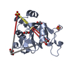
| ||||||||||||
| 3 | 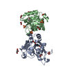
| ||||||||||||
| Unit cell |
|
-Components
| #1: Protein | Mass: 18823.826 Da / Num. of mol.: 2 Source method: isolated from a genetically manipulated source Source: (gene. exp.) Homo sapiens (human) / Gene: YTHDC1, KIAA1966, YT521 / Production host:  #2: Chemical | ChemComp-SO4 /   Mass: 96.063 Da / Num. of mol.: 6 / Source method: obtained synthetically / Formula: SO4 Mass: 96.063 Da / Num. of mol.: 6 / Source method: obtained synthetically / Formula: SO4#3: Chemical | ChemComp-EDO /   Mass: 62.068 Da / Num. of mol.: 8 / Source method: obtained synthetically / Formula: C2H6O2 Mass: 62.068 Da / Num. of mol.: 8 / Source method: obtained synthetically / Formula: C2H6O2#4: Chemical | ChemComp-GOL / |   Mass: 92.094 Da / Num. of mol.: 1 / Source method: obtained synthetically / Formula: C3H8O3 Mass: 92.094 Da / Num. of mol.: 1 / Source method: obtained synthetically / Formula: C3H8O3#5: Water | ChemComp-HOH / |  Mass: 18.015 Da / Num. of mol.: 386 / Source method: isolated from a natural source / Formula: H2O Mass: 18.015 Da / Num. of mol.: 386 / Source method: isolated from a natural source / Formula: H2OHas ligand of interest | N | |
|---|
-Experimental details
-Experiment
| Experiment | Method: X-RAY DIFFRACTION / Number of used crystals: 1 |
|---|
- Sample preparation
| Crystal | Density Matthews: 2.29 Å3/Da / Density % sol: 46.22 % |
|---|---|
| Crystal grow | Temperature: 292 K / Method: vapor diffusion, sitting drop / pH: 5.5 Details: 0.1-0.15M ammonium sulfate 0.1 M Bis-Tris pH 5.5 25-29% PEG 3350 |
-Data collection
| Diffraction | Mean temperature: 100 K / Serial crystal experiment: N |
|---|---|
| Diffraction source | Source: SYNCHROTRON / Site: APS / Beamline: 22-ID / Wavelength: 1 Å |
| Detector | Type: DECTRIS EIGER X 16M / Detector: PIXEL / Date: Dec 19, 2019 |
| Radiation | Protocol: SINGLE WAVELENGTH / Monochromatic (M) / Laue (L): M / Scattering type: x-ray |
| Radiation wavelength | Wavelength: 1 Å / Relative weight: 1 |
| Reflection | Resolution: 1.18→20.01 Å / Num. obs: 93137 / % possible obs: 86.1 % / Redundancy: 5.8 % / Biso Wilson estimate: 11.05 Å2 / Rmerge(I) obs: 0.072 / Rpim(I) all: 0.038 / Net I/σ(I): 22.2 |
| Reflection shell | Resolution: 1.18→1.22 Å / Redundancy: 1.4 % / Rmerge(I) obs: 0.579 / Mean I/σ(I) obs: 1.2 / Num. unique obs: 2859 / CC1/2: 0.41 / CC star: 0.763 / Rpim(I) all: 0.517 / % possible all: 26.5 |
- Processing
| Software |
| |||||||||||||||||||||||||||||||||||||||||||||||||||||||||||||||||||||||||||||||||||||||||||||||||||||||||
|---|---|---|---|---|---|---|---|---|---|---|---|---|---|---|---|---|---|---|---|---|---|---|---|---|---|---|---|---|---|---|---|---|---|---|---|---|---|---|---|---|---|---|---|---|---|---|---|---|---|---|---|---|---|---|---|---|---|---|---|---|---|---|---|---|---|---|---|---|---|---|---|---|---|---|---|---|---|---|---|---|---|---|---|---|---|---|---|---|---|---|---|---|---|---|---|---|---|---|---|---|---|---|---|---|---|---|
| Refinement | Method to determine structure: MOLECULAR REPLACEMENT Starting model: 4r3h Resolution: 1.18→20.01 Å / SU ML: 0.0923 / Cross valid method: FREE R-VALUE / σ(F): 1.35 / Phase error: 22.183 Stereochemistry target values: GeoStd + Monomer Library + CDL v1.2
| |||||||||||||||||||||||||||||||||||||||||||||||||||||||||||||||||||||||||||||||||||||||||||||||||||||||||
| Solvent computation | Shrinkage radii: 0.9 Å / VDW probe radii: 1.11 Å / Solvent model: FLAT BULK SOLVENT MODEL | |||||||||||||||||||||||||||||||||||||||||||||||||||||||||||||||||||||||||||||||||||||||||||||||||||||||||
| Displacement parameters | Biso mean: 20.47 Å2 | |||||||||||||||||||||||||||||||||||||||||||||||||||||||||||||||||||||||||||||||||||||||||||||||||||||||||
| Refinement step | Cycle: LAST / Resolution: 1.18→20.01 Å
| |||||||||||||||||||||||||||||||||||||||||||||||||||||||||||||||||||||||||||||||||||||||||||||||||||||||||
| Refine LS restraints |
| |||||||||||||||||||||||||||||||||||||||||||||||||||||||||||||||||||||||||||||||||||||||||||||||||||||||||
| LS refinement shell |
|
