 Movie
Movie Controller


+ Open data

- Basic information
| Entry | Database: PDB / ID: 4r3h | ||||||
|---|---|---|---|---|---|---|---|
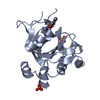
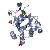
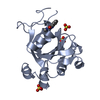
| Title | The crystal structure of an apo RNA binding protein | ||||||
 Components Components | YTH domain-containing protein 1 | ||||||
 Keywords Keywords | RNA BINDING PROTEIN / structural genomics / Structural Genomics Consortium / SGC | ||||||
| Function / homology |  Function and homology information Function and homology informationmRNA splice site recognition / Nuclear RNA decay / N6-methyladenosine-containing RNA reader activity / dosage compensation by inactivation of X chromosome / regulation of mRNA splicing, via spliceosome / post-transcriptional regulation of gene expression / regulation of alternative mRNA splicing, via spliceosome / molecular sequestering activity / mRNA export from nucleus / brown fat cell differentiation ...mRNA splice site recognition / Nuclear RNA decay / N6-methyladenosine-containing RNA reader activity / dosage compensation by inactivation of X chromosome / regulation of mRNA splicing, via spliceosome / post-transcriptional regulation of gene expression / regulation of alternative mRNA splicing, via spliceosome / molecular sequestering activity / mRNA export from nucleus / brown fat cell differentiation / mRNA splicing, via spliceosome / nuclear speck / mRNA binding / RNA binding / nucleoplasm / nucleus Similarity search - Function | ||||||
| Biological species |  Homo sapiens (human) Homo sapiens (human) | ||||||
| Method |  X-RAY DIFFRACTION / MIR / Resolution: 1.9 Å X-RAY DIFFRACTION / MIR / Resolution: 1.9 Å | ||||||
 Authors Authors | Xu, C. / Liu, K. / Tempel, W. / Li, Y. / Bountra, C. / Arrowsmith, C.H. / Edwards, A.M. / Min, J. / Structural Genomics Consortium (SGC) | ||||||
 Citation Citation | Journal: Nat.Chem.Biol. / Year: 2014 Title: Structural basis for selective binding of m(6)A RNA by the YTHDC1 YTH domain. Authors: Xu, C. / Wang, X. / Liu, K. / Roundtree, I.A. / Tempel, W. / Li, Y. / Lu, Z. / He, C. / Min, J. | ||||||
| History |
|
- Structure visualization
| Structure viewer | Molecule: MolmilJmol/JSmol |
|---|
- Downloads & links
-Download
| PDBx/mmCIF format | 4r3h.cif.gz | 136.5 KB | Display | PDBx/mmCIF format |
|---|---|---|---|---|
| PDB format | pdb4r3h.ent.gz | 108.2 KB | Display | PDB format |
| PDBx/mmJSON format | 4r3h.json.gz | Tree view | PDBx/mmJSON format | |
| Others |  Other downloads Other downloads |
-Validation report
| Arichive directory | https://data.pdbj.org/pub/pdb/validation_reports/r3/4r3hftp://data.pdbj.org/pub/pdb/validation_reports/r3/4r3h | HTTPS FTP |
|---|
-Related structure data


-Links
 PDBj
PDBj- Assembly
| Deposited unit | 
| ||||||||
|---|---|---|---|---|---|---|---|---|---|
| 1 | 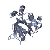
| ||||||||
| 2 | 
| ||||||||
| Unit cell |
|
-Components
| #1: Protein | Mass: 18823.826 Da / Num. of mol.: 2 Source method: isolated from a genetically manipulated source Source: (gene. exp.) Homo sapiens (human) / Gene: YTHDC1, KIAA1966, YT521 / Plasmid: pET28-MHL / Production host:  #2: Chemical | ChemComp-UNX /   Num. of mol.: 11 / Source method: obtained synthetically Num. of mol.: 11 / Source method: obtained synthetically#3: Chemical | ChemComp-SO4 / |   Mass: 96.063 Da / Num. of mol.: 1 / Source method: obtained synthetically / Formula: SO4 Mass: 96.063 Da / Num. of mol.: 1 / Source method: obtained synthetically / Formula: SO4#4: Water | ChemComp-HOH / |  Mass: 18.015 Da / Num. of mol.: 158 / Source method: isolated from a natural source / Formula: H2O Mass: 18.015 Da / Num. of mol.: 158 / Source method: isolated from a natural source / Formula: H2O |
|---|
-Experimental details
-Experiment
| Experiment | Method: X-RAY DIFFRACTION / Number of used crystals: 1 |
|---|
- Sample preparation
| Crystal | Density Matthews: 2.3 Å3/Da / Density % sol: 45.7 % |
|---|---|
| Crystal grow | Temperature: 293 K / Method: vapor diffusion / pH: 6.5 Details: 25% PEG3350, 0.2M Ammonium Sulphate, 0.1M bis-tris, pH 6.5, vapor diffusion, temperature 293K |
-Data collection
| Diffraction | Mean temperature: 100 K | ||||||||||||||||||||||||
|---|---|---|---|---|---|---|---|---|---|---|---|---|---|---|---|---|---|---|---|---|---|---|---|---|---|
| Diffraction source | Source: ROTATING ANODE / Type: RIGAKU / Wavelength: 1.5418 Å | ||||||||||||||||||||||||
| Detector | Type: RIGAKU RAXIS / Detector: IMAGE PLATE / Date: Mar 5, 2014 | ||||||||||||||||||||||||
| Radiation | Protocol: SINGLE WAVELENGTH / Monochromatic (M) / Laue (L): M / Scattering type: x-ray | ||||||||||||||||||||||||
| Radiation wavelength | Wavelength: 1.5418 Å / Relative weight: 1 | ||||||||||||||||||||||||
| Reflection | Resolution: 1.8→27.31 Å / Num. obs: 28931 / % possible obs: 99.1 % / Redundancy: 3.6 % / Rmerge(I) obs: 0.086 / Net I/σ(I): 10.9 | ||||||||||||||||||||||||
| Reflection shell | Diffraction-ID: 1
|
- Processing
| Software |
| |||||||||||||||||||||||||||||||||||||||||||||||||||||||||||||||||||||||||||
|---|---|---|---|---|---|---|---|---|---|---|---|---|---|---|---|---|---|---|---|---|---|---|---|---|---|---|---|---|---|---|---|---|---|---|---|---|---|---|---|---|---|---|---|---|---|---|---|---|---|---|---|---|---|---|---|---|---|---|---|---|---|---|---|---|---|---|---|---|---|---|---|---|---|---|---|---|
| Refinement | Method to determine structure: MIR / Resolution: 1.9→25 Å / Cor.coef. Fo:Fc: 0.955 / Cor.coef. Fo:Fc free: 0.927 / WRfactor Rfree: 0.2194 / WRfactor Rwork: 0.1733 / Occupancy max: 1 / Occupancy min: 0.4 / FOM work R set: 0.8008 / SU B: 8.794 / SU ML: 0.129 / SU R Cruickshank DPI: 0.1619 / SU Rfree: 0.1528 / Cross valid method: THROUGHOUT / σ(F): 0 / ESU R: 0.162 / ESU R Free: 0.153 / Stereochemistry target values: MAXIMUM LIKELIHOOD Details: Structure of an isomorphous crystal was solved by molecular replacement using PHASER and coordinates from PDB entry 2YUD. Phase improvement and automated model building were performed with ...Details: Structure of an isomorphous crystal was solved by molecular replacement using PHASER and coordinates from PDB entry 2YUD. Phase improvement and automated model building were performed with ARP/WARP. COOT was used for interactive model building. Model geometry was evaluated with MOLPROBITY.
| |||||||||||||||||||||||||||||||||||||||||||||||||||||||||||||||||||||||||||
| Solvent computation | Ion probe radii: 0.8 Å / Shrinkage radii: 0.8 Å / VDW probe radii: 1.2 Å / Solvent model: MASK | |||||||||||||||||||||||||||||||||||||||||||||||||||||||||||||||||||||||||||
| Displacement parameters | Biso max: 84.28 Å2 / Biso mean: 32.1817 Å2 / Biso min: 15.45 Å2
| |||||||||||||||||||||||||||||||||||||||||||||||||||||||||||||||||||||||||||
| Refinement step | Cycle: LAST / Resolution: 1.9→25 Å
| |||||||||||||||||||||||||||||||||||||||||||||||||||||||||||||||||||||||||||
| Refine LS restraints |
| |||||||||||||||||||||||||||||||||||||||||||||||||||||||||||||||||||||||||||
| LS refinement shell | Resolution: 1.9→1.949 Å / Total num. of bins used: 20
| |||||||||||||||||||||||||||||||||||||||||||||||||||||||||||||||||||||||||||
| Refinement TLS params. | Method: refined / Refine-ID: X-RAY DIFFRACTION
| |||||||||||||||||||||||||||||||||||||||||||||||||||||||||||||||||||||||||||
| Refinement TLS group |
|
