 Movie
Movie Controller

[English] 日本語
 Yorodumi
- PDB-6zbm: Structure of the D125N mutant of the catalytic domain of the Baci... -
+ Open data

- Basic information
| Entry | Database: PDB / ID: 6zbm | |||||||||
|---|---|---|---|---|---|---|---|---|---|---|
| Title | Structure of the D125N mutant of the catalytic domain of the Bacillus circulans alpha-1,6 Mannanase in complex with an alpha-1,6-alpha-manno-cyclophellitol carbasugar-stabilised trisaccharide inhibitor | |||||||||
 Components Components | Alpha-1,6-mannanase | |||||||||
 Keywords Keywords | HYDROLASE / ENDO-ALPHA-1 / 6-MANNANASE / EPOXIDE / INHIBITOR | |||||||||
| Function / homology |  Function and homology information Function and homology information | |||||||||
| Biological species |  Bacillus circulans (bacteria) Bacillus circulans (bacteria) | |||||||||
| Method |  X-RAY DIFFRACTION / SYNCHROTRON / MOLECULAR REPLACEMENT / Resolution: 1.47 Å X-RAY DIFFRACTION / SYNCHROTRON / MOLECULAR REPLACEMENT / Resolution: 1.47 Å | |||||||||
 Authors Authors | Davies, G.J. / Offen, W.A. | |||||||||
| Funding support |  United Kingdom, 2items United Kingdom, 2items
| |||||||||
 Citation Citation | Journal: Chemistry / Year: 2021 Title: Development of Non-Hydrolysable Oligosaccharide Activity-Based Inactivators for Endoglycanases: A Case Study on alpha-1,6 Mannanases. Authors: Schroder, S.P. / Offen, W.A. / Males, A. / Jin, Y. / de Boer, C. / Enotarpi, J. / Marino, L. / van der Marel, G.A. / Florea, B.I. / Codee, J.D.C. / Overkleeft, H.S. / Davies, G.J. | |||||||||
| History |
|
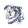
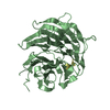
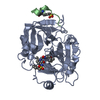
- Structure visualization
| Structure viewer | Molecule: MolmilJmol/JSmol |
|---|
- Downloads & links
-Download
| PDBx/mmCIF format | 6zbm.cif.gz | 158.1 KB | Display | PDBx/mmCIF format |
|---|---|---|---|---|
| PDB format | pdb6zbm.ent.gz | 120.5 KB | Display | PDB format |
| PDBx/mmJSON format | 6zbm.json.gz | Tree view | PDBx/mmJSON format | |
| Others |  Other downloads Other downloads |
-Validation report
| Arichive directory | https://data.pdbj.org/pub/pdb/validation_reports/zb/6zbmftp://data.pdbj.org/pub/pdb/validation_reports/zb/6zbm | HTTPS FTP |
|---|
-Related structure data
| Related structure data |  6zbwC  6zbxC  7nl5C  4d4aS S: Starting model for refinement C: citing same article ( |
|---|---|
| Similar structure data |


-Links
 PDBj
PDBj


- Assembly
| Deposited unit | 
| ||||||||
|---|---|---|---|---|---|---|---|---|---|
| 1 |
| ||||||||
| Unit cell |
|
-Components
| #1: Protein | Mass: 40929.875 Da / Num. of mol.: 1 / Mutation: D125N, R341Q Source method: isolated from a genetically manipulated source Details: Catalytic domain of BcGH76 without signal peptide and with N-terminal His tag, and with mutations D125N and R341Q Source: (gene. exp.) Bacillus circulans (bacteria) / Gene: aman6 / Production host: | ||||||||||
|---|---|---|---|---|---|---|---|---|---|---|---|
| #2: Chemical |   Mass: 62.068 Da / Num. of mol.: 2 / Source method: obtained synthetically / Formula: C2H6O2 Mass: 62.068 Da / Num. of mol.: 2 / Source method: obtained synthetically / Formula: C2H6O2#3: Chemical | ChemComp-QE8 / ( |   Mass: 338.351 Da / Num. of mol.: 1 / Source method: obtained synthetically / Formula: C14H26O9 / Feature type: SUBJECT OF INVESTIGATION Mass: 338.351 Da / Num. of mol.: 1 / Source method: obtained synthetically / Formula: C14H26O9 / Feature type: SUBJECT OF INVESTIGATION#4: Sugar | ChemComp-MAN / |   Type: D-saccharide, alpha linking / Mass: 180.156 Da / Num. of mol.: 1 Type: D-saccharide, alpha linking / Mass: 180.156 Da / Num. of mol.: 1Source method: isolated from a genetically manipulated source Formula: C6H12O6 / Feature type: SUBJECT OF INVESTIGATION #5: Water | ChemComp-HOH / |  Mass: 18.015 Da / Num. of mol.: 178 / Source method: isolated from a natural source / Formula: H2O Mass: 18.015 Da / Num. of mol.: 178 / Source method: isolated from a natural source / Formula: H2OHas ligand of interest | Y | Has protein modification | Y | |
-Experimental details
-Experiment
| Experiment | Method: X-RAY DIFFRACTION / Number of used crystals: 1 |
|---|
- Sample preparation
| Crystal | Density Matthews: 1.72 Å3/Da / Density % sol: 28.43 % |
|---|---|
| Crystal grow | Temperature: 291 K / Method: vapor diffusion, sitting drop / Details: PEG 3350, ammonium nitrate |
-Data collection
| Diffraction | Mean temperature: 100 K / Serial crystal experiment: N |
|---|---|
| Diffraction source | Source: SYNCHROTRON / Site: Diamond / Beamline: I04 / Wavelength: 0.9795 Å |
| Detector | Type: DECTRIS EIGER X 16M / Detector: PIXEL / Date: Jan 18, 2020 |
| Radiation | Protocol: SINGLE WAVELENGTH / Monochromatic (M) / Laue (L): M / Scattering type: x-ray |
| Radiation wavelength | Wavelength: 0.9795 Å / Relative weight: 1 |
| Reflection | Resolution: 1.47→66.12 Å / Num. obs: 47092 / % possible obs: 99.8 % / Redundancy: 4 % / CC1/2: 0.95 / Rpim(I) all: 0.13 / Net I/σ(I): 3.1 |
| Reflection shell | Resolution: 1.47→1.5 Å / Redundancy: 3.4 % / Mean I/σ(I) obs: 1.1 / Num. unique obs: 2333 / CC1/2: 0.676 / Rpim(I) all: 0.631 / % possible all: 99.7 |
- Processing
| Software |
| |||||||||||||||||||||||||||||||||||||||||||||||||||||||||||||||||
|---|---|---|---|---|---|---|---|---|---|---|---|---|---|---|---|---|---|---|---|---|---|---|---|---|---|---|---|---|---|---|---|---|---|---|---|---|---|---|---|---|---|---|---|---|---|---|---|---|---|---|---|---|---|---|---|---|---|---|---|---|---|---|---|---|---|---|
| Refinement | Method to determine structure: MOLECULAR REPLACEMENT Starting model: 4D4A.PDB Resolution: 1.47→48.66 Å / Cor.coef. Fo:Fc: 0.965 / Cor.coef. Fo:Fc free: 0.935 / SU B: 5.101 / SU ML: 0.081 / Cross valid method: THROUGHOUT / σ(F): 0 / ESU R: 0.094 / ESU R Free: 0.083 / Stereochemistry target values: MAXIMUM LIKELIHOOD Details: HYDROGENS HAVE BEEN ADDED IN THE RIDING POSITIONS U VALUES : REFINED INDIVIDUALLY THERE ARE REGIONS OF UNMODELLED DENSITY NEAR THE SIDE CHAINS OF ASN206, HIS340 AND TYR360.
| |||||||||||||||||||||||||||||||||||||||||||||||||||||||||||||||||
| Solvent computation | Ion probe radii: 0.8 Å / Shrinkage radii: 0.8 Å / VDW probe radii: 1.2 Å / Solvent model: MASK | |||||||||||||||||||||||||||||||||||||||||||||||||||||||||||||||||
| Displacement parameters | Biso max: 52.29 Å2 / Biso mean: 13.884 Å2 / Biso min: 6.74 Å2
| |||||||||||||||||||||||||||||||||||||||||||||||||||||||||||||||||
| Refinement step | Cycle: final / Resolution: 1.47→48.66 Å
| |||||||||||||||||||||||||||||||||||||||||||||||||||||||||||||||||
| Refine LS restraints |
| |||||||||||||||||||||||||||||||||||||||||||||||||||||||||||||||||
| LS refinement shell | Resolution: 1.47→1.508 Å / Rfactor Rfree error: 0 / Total num. of bins used: 20
|
