 Movie
Movie Controller

[English] 日本語
 Yorodumi
- PDB-6zb8: Exo-beta-1,3-glucanase from moose rumen microbiome, active site m... -
+ Open data

- Basic information
| Entry | Database: PDB / ID: 6zb8 | ||||||
|---|---|---|---|---|---|---|---|
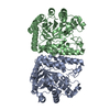
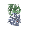
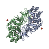
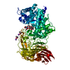
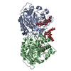
| Title | Exo-beta-1,3-glucanase from moose rumen microbiome, active site mutant E167Q/E295Q | ||||||
 Components Components | Exo-beta-1,3-glucanase variant E167Q/E295Q | ||||||
 Keywords Keywords | HYDROLASE / Glycoside hydrolase / Exo-beta-1 / 3-glucanase / family GH5_44 / active-site mutant | ||||||
| Function / homology | Glycosidases / TIM Barrel / Alpha-Beta Barrel / Alpha Beta Function and homology information Function and homology information | ||||||
| Biological species |  uncultured bacterium (environmental samples) uncultured bacterium (environmental samples) | ||||||
| Method |  X-RAY DIFFRACTION / SYNCHROTRON / FOURIER SYNTHESIS / Resolution: 1.35 Å X-RAY DIFFRACTION / SYNCHROTRON / FOURIER SYNTHESIS / Resolution: 1.35 Å | ||||||
 Authors Authors | Kalyani, D.C. / Reichenbach, T. / Aspeborg, H. / Divne, C. | ||||||
| Funding support |  Sweden, 1items Sweden, 1items
| ||||||
 Citation Citation | Journal: Enzyme.Microb.Technol. / Year: 2021 Title: A homodimeric bacterial exo-beta-1,3-glucanase derived from moose rumen microbiome shows a structural framework similar to yeast exo-beta-1,3-glucanases. Authors: Kalyani, D.C. / Reichenbach, T. / Aspeborg, H. / Divne, C. | ||||||
| History |
|
- Structure visualization
| Structure viewer | Molecule: MolmilJmol/JSmol |
|---|
- Downloads & links
-Download
| PDBx/mmCIF format | 6zb8.cif.gz | 307.9 KB | Display | PDBx/mmCIF format |
|---|---|---|---|---|
| PDB format | pdb6zb8.ent.gz | 248.8 KB | Display | PDB format |
| PDBx/mmJSON format | 6zb8.json.gz | Tree view | PDBx/mmJSON format | |
| Others |  Other downloads Other downloads |
-Validation report
| Summary document | 6zb8_validation.pdf.gz | 771.7 KB | Display | wwPDB validaton report |
|---|---|---|---|---|
| Full document | 6zb8_full_validation.pdf.gz | 775.4 KB | Display | |
| Data in XML | 6zb8_validation.xml.gz | 33 KB | Display | |
| Data in CIF | 6zb8_validation.cif.gz | 49.7 KB | Display | |
| Arichive directory | https://data.pdbj.org/pub/pdb/validation_reports/zb/6zb8ftp://data.pdbj.org/pub/pdb/validation_reports/zb/6zb8 | HTTPS FTP |
-Related structure data


-Links
 PDBj
PDBj- Assembly
| Deposited unit | 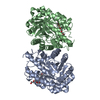
| ||||||||
|---|---|---|---|---|---|---|---|---|---|
| 1 |
| ||||||||
| Unit cell |
|
-Components
| #1: Protein | Mass: 45379.711 Da / Num. of mol.: 2 / Mutation: E167Q, E295Q Source method: isolated from a genetically manipulated source Details: An active-site loop is disordered in the model and has therefore not been modeled. Source: (gene. exp.) uncultured bacterium (environmental samples)Plasmid: pET-11a / Production host: #2: Chemical |   Mass: 1529.829 Da / Num. of mol.: 2 / Source method: obtained synthetically / Formula: C69H140O35 / Comment: precipitant*YM Mass: 1529.829 Da / Num. of mol.: 2 / Source method: obtained synthetically / Formula: C69H140O35 / Comment: precipitant*YM#3: Water | ChemComp-HOH / |  Mass: 18.015 Da / Num. of mol.: 624 / Source method: isolated from a natural source / Formula: H2O Mass: 18.015 Da / Num. of mol.: 624 / Source method: isolated from a natural source / Formula: H2OHas ligand of interest | N | |
|---|
-Experimental details
-Experiment
| Experiment | Method: X-RAY DIFFRACTION / Number of used crystals: 1 |
|---|
- Sample preparation
| Crystal | Density Matthews: 1.97 Å3/Da / Density % sol: 42 % |
|---|---|
| Crystal grow | Temperature: 298 K / Method: vapor diffusion, hanging drop / pH: 8.5 / Details: 0.1M TrisHCl pH 8.5, 0.2M MgCl, 30% PEG 4000 |
-Data collection
| Diffraction | Mean temperature: 100 K / Serial crystal experiment: N |
|---|---|
| Diffraction source | Source: SYNCHROTRON / Site: MAX IV / Beamline: BioMAX / Wavelength: 0.826561 Å |
| Detector | Type: DECTRIS EIGER X 16M / Detector: PIXEL / Date: Mar 27, 2019 / Details: Kirkpatrick-Baez mirror pair |
| Radiation | Monochromator: Si (111), double crystal monochromator / Protocol: SINGLE WAVELENGTH / Monochromatic (M) / Laue (L): M / Scattering type: x-ray |
| Radiation wavelength | Wavelength: 0.826561 Å / Relative weight: 1 |
| Reflection | Resolution: 1.35→29.35 Å / Num. obs: 139214 / % possible obs: 95.2 % / Redundancy: 6.6 % / CC1/2: 0.997 / Rsym value: 0.084 / Net I/σ(I): 13.2 |
| Reflection shell | Resolution: 1.35→1.4 Å / Redundancy: 5 % / Mean I/σ(I) obs: 1.1 / Num. unique obs: 10636 / CC1/2: 0.373 / % possible all: 70.8 |
- Processing
| Software |
| ||||||||||||||||||||||||||||||||||||||||||||||||||||||||||||||||||||||||||||||||||||||||||||||||||
|---|---|---|---|---|---|---|---|---|---|---|---|---|---|---|---|---|---|---|---|---|---|---|---|---|---|---|---|---|---|---|---|---|---|---|---|---|---|---|---|---|---|---|---|---|---|---|---|---|---|---|---|---|---|---|---|---|---|---|---|---|---|---|---|---|---|---|---|---|---|---|---|---|---|---|---|---|---|---|---|---|---|---|---|---|---|---|---|---|---|---|---|---|---|---|---|---|---|---|---|
| Refinement | Method to determine structure: FOURIER SYNTHESIS Starting model: Wild-type structure solved by MR-SAD Resolution: 1.35→29.35 Å / SU ML: 0.14 / Cross valid method: FREE R-VALUE / σ(F): 1.36 / Phase error: 18.86
| ||||||||||||||||||||||||||||||||||||||||||||||||||||||||||||||||||||||||||||||||||||||||||||||||||
| Solvent computation | Shrinkage radii: 0.9 Å / VDW probe radii: 1.11 Å | ||||||||||||||||||||||||||||||||||||||||||||||||||||||||||||||||||||||||||||||||||||||||||||||||||
| Refinement step | Cycle: LAST / Resolution: 1.35→29.35 Å
| ||||||||||||||||||||||||||||||||||||||||||||||||||||||||||||||||||||||||||||||||||||||||||||||||||
| Refine LS restraints |
| ||||||||||||||||||||||||||||||||||||||||||||||||||||||||||||||||||||||||||||||||||||||||||||||||||
| LS refinement shell |
|
