Movie
Movie Controller

[English] 日本語
 Yorodumi
- PDB-6z5j: Arrangement of the matrix protein M1 in influenza A/Hong Kong/1/1... -
+ Open data

- Basic information
| Entry | Database: PDB / ID: 6z5j | ||||||||||||
|---|---|---|---|---|---|---|---|---|---|---|---|---|---|
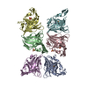
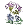
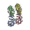
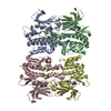
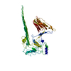
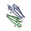
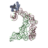
| Title | Arrangement of the matrix protein M1 in influenza A/Hong Kong/1/1968 VLPs (HA,NA,M1,M2) | ||||||||||||
 Components Components | Matrix protein 1 | ||||||||||||
 Keywords Keywords | VIRAL PROTEIN / Viral matrix protein / Membrane binding / pH Sensor / Polymer | ||||||||||||
| Function / homology |  Function and homology information Function and homology informationvirion assembly / viral budding from plasma membrane / structural constituent of virion / host cell nucleus / virion membrane / RNA binding Similarity search - Function | ||||||||||||
| Biological species |   Influenza A virus Influenza A virus | ||||||||||||
| Method | ELECTRON MICROSCOPY / subtomogram averaging / cryo EM / Resolution: 8 Å | ||||||||||||
 Authors Authors | Peukes, J. / Xiong, X. / Erlendsson, S. / Qu, K. / Wan, W. / Kraeusslich, H.-G. / Briggs, J.A.G. | ||||||||||||
| Funding support | European Union,  United Kingdom, United Kingdom,  Germany, 3items Germany, 3items
| ||||||||||||
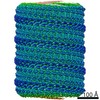
 Citation Citation | Journal: Nature / Year: 2020 Title: The native structure of the assembled matrix protein 1 of influenza A virus. Authors: Julia Peukes / Xiaoli Xiong / Simon Erlendsson / Kun Qu / William Wan / Leslie J Calder / Oliver Schraidt / Susann Kummer / Stefan M V Freund / Hans-Georg Kräusslich / John A G Briggs /    Abstract: Influenza A virus causes millions of severe cases of disease during annual epidemics. The most abundant protein in influenza virions is matrix protein 1 (M1), which mediates virus assembly by ...Influenza A virus causes millions of severe cases of disease during annual epidemics. The most abundant protein in influenza virions is matrix protein 1 (M1), which mediates virus assembly by forming an endoskeleton beneath the virus membrane. The structure of full-length M1, and how it oligomerizes to mediate the assembly of virions, is unknown. Here we determine the complete structure of assembled M1 within intact virus particles, as well as the structure of M1 oligomers reconstituted in vitro. We find that the C-terminal domain of M1 is disordered in solution but can fold and bind in trans to the N-terminal domain of another M1 monomer, thus polymerizing M1 into linear strands that coat the interior surface of the membrane of the assembling virion. In the M1 polymer, five histidine residues-contributed by three different monomers of M1-form a cluster that can serve as the pH-sensitive disassembly switch after entry into a target cell. These structures therefore reveal mechanisms of influenza virus assembly and disassembly. | ||||||||||||
| History |
|
- Structure visualization
| Movie |
Movie viewer |
|---|---|
| Structure viewer | Molecule: MolmilJmol/JSmol |
- Downloads & links
-Download
| PDBx/mmCIF format | 6z5j.cif.gz | 197.4 KB | Display | PDBx/mmCIF format |
|---|---|---|---|---|
| PDB format | pdb6z5j.ent.gz | 154.7 KB | Display | PDB format |
| PDBx/mmJSON format | 6z5j.json.gz | Tree view | PDBx/mmJSON format | |
| Others |  Other downloads Other downloads |
-Validation report
| Arichive directory | https://data.pdbj.org/pub/pdb/validation_reports/z5/6z5jftp://data.pdbj.org/pub/pdb/validation_reports/z5/6z5j | HTTPS FTP |
|---|
-Related structure data
| Related structure data |  11078MC 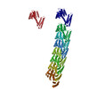 6z5lC M: map data used to model this data C: citing same article ( |
|---|---|
| Similar structure data |

-Links
 PDBj
PDBj

- Assembly
| Deposited unit | 
|
|---|---|
| 1 |
|
-Components
| #1: Protein | Mass: 27928.301 Da / Num. of mol.: 6 Source method: isolated from a genetically manipulated source Details: The PDB model was generated by rigid body fitting of the M1 NTD crystal structure (PBD: 1ea3, from PR8 influenza virus) into the EM density map obtained for HK68 VLPs. Source: (gene. exp.) Influenza A virus (A/Puerto Rico/8-9NMC3/1934(H1N1))Strain: A/Puerto Rico/8-9NMC3/1934(H1N1) / Gene: M1, M / Cell line (production host): HEK293-T / Production host:  Homo sapiens (human) / References: UniProt: F0TTD6 Homo sapiens (human) / References: UniProt: F0TTD6#2: Water | ChemComp-HOH / |  Mass: 18.015 Da / Num. of mol.: 258 / Source method: isolated from a natural source / Formula: H2O Mass: 18.015 Da / Num. of mol.: 258 / Source method: isolated from a natural source / Formula: H2O |
|---|
-Experimental details
-Experiment
| Experiment | Method: ELECTRON MICROSCOPY |
|---|---|
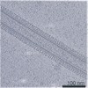
| EM experiment | Aggregation state: PARTICLE / 3D reconstruction method: subtomogram averaging |
- Sample preparation
| Component |
| |||||||||||||||||||||
|---|---|---|---|---|---|---|---|---|---|---|---|---|---|---|---|---|---|---|---|---|---|---|
| Source (natural) |
| |||||||||||||||||||||
| Source (recombinant) |
| |||||||||||||||||||||
| Details of virus |
| |||||||||||||||||||||
| Natural host |
| |||||||||||||||||||||
| Buffer solution | pH: 7.5 | |||||||||||||||||||||
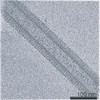
| Specimen | Embedding applied: NO / Shadowing applied: NO / Staining applied: NO / Vitrification applied: YES | |||||||||||||||||||||
| Specimen support | Grid material: GOLD / Grid mesh size: 200 divisions/in. / Grid type: Quantifoil R2/2 | |||||||||||||||||||||
| Vitrification | Instrument: LEICA EM GP / Cryogen name: ETHANE / Humidity: 95 % |
- Electron microscopy imaging
| Experimental equipment |  Model: Titan Krios / Image courtesy: FEI Company |
|---|---|
| Microscopy | Model: FEI TITAN KRIOS |
| Electron gun | Electron source:  FIELD EMISSION GUN / Accelerating voltage: 300 kV / Illumination mode: FLOOD BEAM FIELD EMISSION GUN / Accelerating voltage: 300 kV / Illumination mode: FLOOD BEAM |
| Electron lens | Mode: BRIGHT FIELD / Nominal magnification: 81000 X / Nominal defocus max: 4000 nm / Nominal defocus min: 2000 nm / Cs: 2.7 mm / Alignment procedure: ZEMLIN TABLEAU |
| Specimen holder | Cryogen: NITROGEN / Specimen holder model: FEI TITAN KRIOS AUTOGRID HOLDER |
| Image recording | Electron dose: 2.9 e/Å2 / Detector mode: SUPER-RESOLUTION / Film or detector model: GATAN K2 QUANTUM (4k x 4k) |
| EM imaging optics | Energyfilter name: GIF Quantum LS / Energyfilter slit width: 20 eV |
| Image scans | Movie frames/image: 20 |
- Processing
| EM software |
| |||||||||||||||||||||||||||
|---|---|---|---|---|---|---|---|---|---|---|---|---|---|---|---|---|---|---|---|---|---|---|---|---|---|---|---|---|
| CTF correction | Details: CTF determination was performed using CTFFIND4. CTF correction was performed by 3D-CTF correction by CTF multiplication in NovaCTF. Type: PHASE FLIPPING AND AMPLITUDE CORRECTION | |||||||||||||||||||||||||||
| Symmetry | Point symmetry: C1 (asymmetric) | |||||||||||||||||||||||||||
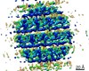
| 3D reconstruction | Resolution: 8 Å / Resolution method: FSC 0.143 CUT-OFF / Num. of particles: 14767 / Algorithm: BACK PROJECTION / Symmetry type: POINT | |||||||||||||||||||||||||||
| EM volume selection | Method: Volumes picked semi-automatically along the particles membrane Details: Initial positions were generated along the surface of a cylinder simulated around the manually determined central axis of each particle. Initial positions were at least 2X oversampled. Num. of tomograms: 4 / Num. of volumes extracted: 120000 | |||||||||||||||||||||||||||
| Atomic model building | Protocol: RIGID BODY FIT Details: Multiple copies of the M1 NTD crystal structure (PDB:1ea3) were fitted as rigid bodies into the EM map to understand the relative arrangement of M1 monomers in the context of the matrix layer inside the virus | |||||||||||||||||||||||||||
| Atomic model building | PDB-ID: 1EA3 Pdb chain-ID: A / Accession code: 1EA3 / Source name: PDB / Type: experimental model |