 Movie
Movie Controller

[English] 日本語
 Yorodumi
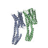
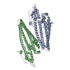
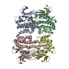
- PDB-5ly6: CryoEM structure of the membrane pore complex of Pneumolysin at 4.5A -
+ Open data

- Basic information
| Entry | Database: PDB / ID: 5ly6 | ||||||
|---|---|---|---|---|---|---|---|
| Title | CryoEM structure of the membrane pore complex of Pneumolysin at 4.5A | ||||||
 Components Components | Pneumolysin | ||||||
 Keywords Keywords | TOXIN / Bacterial toxins / pore complex / cryoEM structure / cholesterol-dependent cytolysin / Pneumolysin / membrane pore | ||||||
| Function / homology |  Function and homology information Function and homology informationcholesterol binding / toxin activity / killing of cells of another organism / host cell plasma membrane / extracellular region Similarity search - Function | ||||||
| Biological species | Streptococcus pneumoniae serotype 2 | ||||||
| Method | ELECTRON MICROSCOPY / single particle reconstruction / cryo EM / Resolution: 4.5 Å | ||||||
 Authors Authors | van Pee, K. / Neuhaus, A. / D'Imprima, E. / Mills, D.J. / Kuehlbrandt, W. / Yildiz, O. | ||||||
| Funding support |  Germany, 1items Germany, 1items
| ||||||
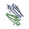
 Citation Citation | Journal: Elife / Year: 2017 Title: CryoEM structures of membrane pore and prepore complex reveal cytolytic mechanism of Pneumolysin. Authors: Katharina van Pee / Alexander Neuhaus / Edoardo D'Imprima / Deryck J Mills / Werner Kühlbrandt / Özkan Yildiz / Abstract: Many pathogenic bacteria produce pore-forming toxins to attack and kill human cells. We have determined the 4.5 Å structure of the ~2.2 MDa pore complex of pneumolysin, the main virulence factor of ...Many pathogenic bacteria produce pore-forming toxins to attack and kill human cells. We have determined the 4.5 Å structure of the ~2.2 MDa pore complex of pneumolysin, the main virulence factor of , by cryoEM. The pneumolysin pore is a 400 Å ring of 42 membrane-inserted monomers. Domain 3 of the soluble toxin refolds into two ~85 Å β-hairpins that traverse the lipid bilayer and assemble into a 168-strand β-barrel. The pore complex is stabilized by salt bridges between β-hairpins of adjacent subunits and an internal α-barrel. The apolar outer barrel surface with large sidechains is immersed in the lipid bilayer, while the inner barrel surface is highly charged. Comparison of the cryoEM pore complex to the prepore structure obtained by electron cryo-tomography and the x-ray structure of the soluble form reveals the detailed mechanisms by which the toxin monomers insert into the lipid bilayer to perforate the target membrane. | ||||||
| History |
|
- Structure visualization
| Movie |
Movie viewer |
|---|---|
| Structure viewer | Molecule: MolmilJmol/JSmol |
- Downloads & links
-Download
| PDBx/mmCIF format | 5ly6.cif.gz | 105.9 KB | Display | PDBx/mmCIF format |
|---|---|---|---|---|
| PDB format | pdb5ly6.ent.gz | 80.6 KB | Display | PDB format |
| PDBx/mmJSON format | 5ly6.json.gz | Tree view | PDBx/mmJSON format | |
| Others |  Other downloads Other downloads |
-Validation report
| Arichive directory | https://data.pdbj.org/pub/pdb/validation_reports/ly/5ly6ftp://data.pdbj.org/pub/pdb/validation_reports/ly/5ly6 | HTTPS FTP |
|---|
-Related structure data
| Related structure data |  4118MC  5aoeC 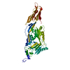 5aofC M: map data used to model this data C: citing same article ( |
|---|---|
| Similar structure data |


-Links
 PDBj
PDBj- Assembly
| Deposited unit | 
|
|---|---|
| 1 |
|
-Components
| #1: Protein | Mass: 52866.066 Da / Num. of mol.: 1 Source method: isolated from a genetically manipulated source Source: (gene. exp.)  Streptococcus pneumoniae serotype 2 (strain D39 / NCTC 7466) (bacteria) Streptococcus pneumoniae serotype 2 (strain D39 / NCTC 7466) (bacteria)Gene: ply, SPD_1726 / Production host: |
|---|
-Experimental details
-Experiment
| Experiment | Method: ELECTRON MICROSCOPY |
|---|---|
| EM experiment | Aggregation state: PARTICLE / 3D reconstruction method: single particle reconstruction |
- Sample preparation
| Component | Name: Pneumolysin pore complex / Type: ORGANELLE OR CELLULAR COMPONENT / Entity ID: all / Source: RECOMBINANT |
|---|---|
| Molecular weight | Value: 2.2 MDa |
| Source (natural) | Organism:  Streptococcus pneumoniae (bacteria) Streptococcus pneumoniae (bacteria) |
| Source (recombinant) | Organism: |
| Buffer solution | pH: 7 |
| Specimen | Conc.: 1 mg/ml / Embedding applied: NO / Shadowing applied: NO / Staining applied: NO / Vitrification applied: YES |
| Vitrification | Cryogen name: ETHANE |
- Electron microscopy imaging
| Experimental equipment |  Model: Tecnai Polara / Image courtesy: FEI Company |
|---|---|
| Microscopy | Model: FEI POLARA 300 |
| Electron gun | Electron source:  FIELD EMISSION GUN / Accelerating voltage: 300 kV / Illumination mode: FLOOD BEAM FIELD EMISSION GUN / Accelerating voltage: 300 kV / Illumination mode: FLOOD BEAM |
| Electron lens | Mode: BRIGHT FIELD |
| Image recording | Average exposure time: 6 sec. / Electron dose: 1.02 e/Å2 / Detector mode: COUNTING / Film or detector model: GATAN K2 SUMMIT (4k x 4k) |
- Processing
| EM software |
| ||||||||||||||||||||||||||||||||||||||||
|---|---|---|---|---|---|---|---|---|---|---|---|---|---|---|---|---|---|---|---|---|---|---|---|---|---|---|---|---|---|---|---|---|---|---|---|---|---|---|---|---|---|
| CTF correction | Type: PHASE FLIPPING AND AMPLITUDE CORRECTION | ||||||||||||||||||||||||||||||||||||||||
| Symmetry | Point symmetry: C42 (42 fold cyclic) | ||||||||||||||||||||||||||||||||||||||||
| 3D reconstruction | Resolution: 4.5 Å / Resolution method: FSC 0.143 CUT-OFF / Num. of particles: 6461 / Symmetry type: POINT | ||||||||||||||||||||||||||||||||||||||||
| Atomic model building | Protocol: AB INITIO MODEL | ||||||||||||||||||||||||||||||||||||||||
| Atomic model building | PDB-ID: 5AOD Pdb chain-ID: A / Accession code: 5AOD / Pdb chain residue range: 1-471 / Source name: PDB / Type: experimental model |

