 Movie
Movie Controller

[English] 日本語
 Yorodumi
- PDB-6vbs: The C2 Crystal form of SodCI Superoxide Dismutase at 1.7 A resolu... -
+ Open data

- Basic information
| Entry | Database: PDB / ID: 6vbs | ||||||
|---|---|---|---|---|---|---|---|
| Title | The C2 Crystal form of SodCI Superoxide Dismutase at 1.7 A resolution with 6 molecules in the asymmetric unit. | ||||||
 Components Components | Superoxide dismutase [Cu-Zn] | ||||||
 Keywords Keywords | OXYGEN BINDING / Superoxide Dismutase / virulence factor / peptidoglycan binding | ||||||
| Function / homology |  Function and homology information Function and homology informationsuperoxide dismutase / superoxide dismutase activity / removal of superoxide radicals / periplasmic space / copper ion binding / identical protein binding Similarity search - Function | ||||||
| Biological species |  Salmonella typhimurium (bacteria) Salmonella typhimurium (bacteria) | ||||||
| Method |  X-RAY DIFFRACTION / SYNCHROTRON / MOLECULAR REPLACEMENT / Resolution: 1.7 Å X-RAY DIFFRACTION / SYNCHROTRON / MOLECULAR REPLACEMENT / Resolution: 1.7 Å | ||||||
 Authors Authors | Satyshur, K.A. / Forest, K.T. / Newhouse, P.W. | ||||||
| Funding support |  United States, 1items United States, 1items
| ||||||
 Citation Citation | Journal: To Be Published Title: Structure and Muropeptide Binding of the Virulence Factor Superoxide Dismutase C1 from Salmonella Typhimurium Authors: Newhouse, P.W. / Satyshur, K.A. / Slauch, J.M. / Forest, K.T. | ||||||
| History |
|
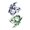
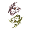
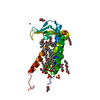
- Structure visualization
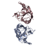
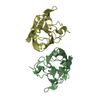
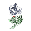
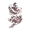
| Structure viewer | Molecule: MolmilJmol/JSmol |
|---|
- Downloads & links
-Download
| PDBx/mmCIF format | 6vbs.cif.gz | 350.4 KB | Display | PDBx/mmCIF format |
|---|---|---|---|---|
| PDB format | pdb6vbs.ent.gz | 284.5 KB | Display | PDB format |
| PDBx/mmJSON format | 6vbs.json.gz | Tree view | PDBx/mmJSON format | |
| Others |  Other downloads Other downloads |
-Validation report
| Arichive directory | https://data.pdbj.org/pub/pdb/validation_reports/vb/6vbsftp://data.pdbj.org/pub/pdb/validation_reports/vb/6vbs | HTTPS FTP |
|---|
-Related structure data
| Related structure data | 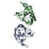 6vbtC  6d52S S: Starting model for refinement C: citing same article ( |
|---|---|
| Similar structure data |
-Links
 PDBj
PDBj


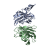
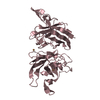
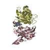
- Assembly
| Deposited unit | 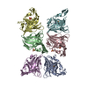
| ||||||||||||
|---|---|---|---|---|---|---|---|---|---|---|---|---|---|
| 1 | 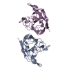
| ||||||||||||
| 2 | 
| ||||||||||||
| 3 | 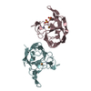
| ||||||||||||
| Unit cell |
| ||||||||||||
| Components on special symmetry positions |
|
-Components
| #1: Protein | Mass: 17437.734 Da / Num. of mol.: 6 Source method: isolated from a genetically manipulated source Source: (gene. exp.) Salmonella typhimurium (bacteria)Gene: sodC1, AAP89_24585, ABO94_13165, AF480_24525, AF488_17800, AF489_22535, AIC76_23135, AU613_25900, AXR84_22600, AXU58_25005, C2253_23510, CD48_19550, CE87_24875, CET98_19490, CQG18_24025, CVR97_ ...Gene: sodC1, AAP89_24585, ABO94_13165, AF480_24525, AF488_17800, AF489_22535, AIC76_23135, AU613_25900, AXR84_22600, AXU58_25005, C2253_23510, CD48_19550, CE87_24875, CET98_19490, CQG18_24025, CVR97_00185, D4369_24860, D4380_23405, D4401_24550, D4E62_23815, D6360_24640, D7F20_18080, D7H43_24310, DD95_21320, DJ388_22940, DKJ11_15870, DKU57_23325, DLM31_21285, DO698_19845, DOJ90_22365, DOQ88_23710, DQ848_24435, DRM14_23695, DSF69_10260, DSR36_22945, DUW48_23400, EBO41_23675, EBP31_23480, EGU67_12495, EHB24_23490, EHC98_23755, EIW53_22710, F0A00_10055, F0A01_03930, F0A02_16630, F0A03_14260, F0A04_11295, F0A05_20695, F0A06_10485, F0U66_16995, FKA80_23505, FKA81_22545, FKA82_23465, FKA83_23450, FKA84_23540, FKA85_22415, FKA86_23320, FKA87_22565, FKA88_23655, FKA89_19440, FKA90_23025, FKA91_22685, FKA92_23550, FKA93_23530, FKA94_23705, FKA95_23590, FKA96_23695, FKA97_23460, FKA98_23465, FKA99_23670, FKB00_23595, FKB01_23750, FKB02_23725, FKB03_23595, FKB12_23280, FKB15_23500, FKB18_22510, FKB19_23085, FYL57_20425, FYL66_21285, FYL68_13470, FYL69_02095, FYL71_08605, FYL73_12165, FYL74_21170, FYL75_22020, FYL77_03645, FYL81_07750, FYL84_20475, FYL90_20575, FYL92_04365, FYL93_18435, FYL94_21135, FYL96_21895, FYM06_15765, FYM08_18765, FYM09_02430, FYM54_04305, FZ992_22195, FZ993_21050, FZ994_04265, FZ995_00435, FZ996_12505, FZ997_14980, FZ998_14695, FZ999_05915, GW08_24555, JO10_24570, LZ63_20420, NCTC13348_01621, NG18_23390, NU83_09530, QA89_23615, QD15_24180, RJ78_24990, SAMEA4398682_02945, Y934_24990, YG50_23900, YI33_24375, YR17_24730, ZB89_24710, ZC54_24870 Production host: References: UniProt: A0A0D6GQL3, UniProt: P0CW86*PLUS, superoxide dismutase #2: Chemical | ChemComp-CU /   Mass: 63.546 Da / Num. of mol.: 6 / Source method: obtained synthetically / Formula: Cu / Feature type: SUBJECT OF INVESTIGATION Mass: 63.546 Da / Num. of mol.: 6 / Source method: obtained synthetically / Formula: Cu / Feature type: SUBJECT OF INVESTIGATION#3: Chemical | ChemComp-ZN /   Mass: 65.409 Da / Num. of mol.: 6 / Source method: obtained synthetically / Formula: Zn / Feature type: SUBJECT OF INVESTIGATION Mass: 65.409 Da / Num. of mol.: 6 / Source method: obtained synthetically / Formula: Zn / Feature type: SUBJECT OF INVESTIGATION#4: Chemical | ChemComp-SO4 /   Mass: 96.063 Da / Num. of mol.: 4 / Source method: obtained synthetically / Formula: SO4 Mass: 96.063 Da / Num. of mol.: 4 / Source method: obtained synthetically / Formula: SO4#5: Water | ChemComp-HOH / |  Mass: 18.015 Da / Num. of mol.: 849 / Source method: isolated from a natural source / Formula: H2O Mass: 18.015 Da / Num. of mol.: 849 / Source method: isolated from a natural source / Formula: H2OHas ligand of interest | Y | Has protein modification | Y | |
|---|
-Experimental details
-Experiment
| Experiment | Method: X-RAY DIFFRACTION / Number of used crystals: 1 |
|---|
- Sample preparation
| Crystal | Density Matthews: 2.16 Å3/Da / Density % sol: 42.96 % |
|---|---|
| Crystal grow | Temperature: 300 K / Method: vapor diffusion, hanging drop / pH: 6.5 Details: 20 mg/ml SODC1 plus 1.2 mM trachaeal cytotoxin in 20 mM Tris pH 6.6 150 mM NaCl set up as hanging drop with mother liquor Bis-Tris 0.05M, Ammonium Sulfate 0.05M, 30% Pentaerythritol ethoxylate (15/4 EO/OH) |
-Data collection
| Diffraction | Mean temperature: 100 K / Serial crystal experiment: N |
|---|---|
| Diffraction source | Source: SYNCHROTRON / Site: APS / Beamline: 21-ID-D / Wavelength: 1.07812 Å |
| Detector | Type: DECTRIS EIGER X 9M / Detector: PIXEL / Date: May 30, 2019 |
| Radiation | Protocol: SINGLE WAVELENGTH / Monochromatic (M) / Laue (L): M / Scattering type: x-ray |
| Radiation wavelength | Wavelength: 1.07812 Å / Relative weight: 1 |
| Reflection | Resolution: 1.528→84.987 Å / Num. obs: 187907 / % possible obs: 79.5 % / Redundancy: 6.6 % / Biso Wilson estimate: 19.48 Å2 / CC1/2: 0.998 / Rmerge(I) obs: 0.062 / Rpim(I) all: 0.026 / Rrim(I) all: 0.067 / Net I/σ(I): 14.4 |
| Reflection shell | Resolution: 1.528→1.639 Å / Redundancy: 4.9 % / Rmerge(I) obs: 0.598 / Mean I/σ(I) obs: 1.7 / Num. unique obs: 5352 / CC1/2: 0.768 / Rpim(I) all: 0.297 / Rrim(I) all: 0.671 / % possible all: 21 |
- Processing
| Software |
| ||||||||||||||||||||||||||||||||||||||||||||||||||||||||||||||||||||||||||||||||||||||||||||||||||||||||||||||||||||||||||||||||||||||||||||||||||||||||||||||||||||||||||||||||||||||
|---|---|---|---|---|---|---|---|---|---|---|---|---|---|---|---|---|---|---|---|---|---|---|---|---|---|---|---|---|---|---|---|---|---|---|---|---|---|---|---|---|---|---|---|---|---|---|---|---|---|---|---|---|---|---|---|---|---|---|---|---|---|---|---|---|---|---|---|---|---|---|---|---|---|---|---|---|---|---|---|---|---|---|---|---|---|---|---|---|---|---|---|---|---|---|---|---|---|---|---|---|---|---|---|---|---|---|---|---|---|---|---|---|---|---|---|---|---|---|---|---|---|---|---|---|---|---|---|---|---|---|---|---|---|---|---|---|---|---|---|---|---|---|---|---|---|---|---|---|---|---|---|---|---|---|---|---|---|---|---|---|---|---|---|---|---|---|---|---|---|---|---|---|---|---|---|---|---|---|---|---|---|---|---|
| Refinement | Method to determine structure: MOLECULAR REPLACEMENT Starting model: 6D52 Resolution: 1.7→35.88 Å / SU ML: 0.178 / Cross valid method: FREE R-VALUE / σ(F): 1.39 / Phase error: 21.1984
| ||||||||||||||||||||||||||||||||||||||||||||||||||||||||||||||||||||||||||||||||||||||||||||||||||||||||||||||||||||||||||||||||||||||||||||||||||||||||||||||||||||||||||||||||||||||
| Solvent computation | Shrinkage radii: 0.9 Å / VDW probe radii: 1.11 Å | ||||||||||||||||||||||||||||||||||||||||||||||||||||||||||||||||||||||||||||||||||||||||||||||||||||||||||||||||||||||||||||||||||||||||||||||||||||||||||||||||||||||||||||||||||||||
| Displacement parameters | Biso mean: 24.1 Å2 | ||||||||||||||||||||||||||||||||||||||||||||||||||||||||||||||||||||||||||||||||||||||||||||||||||||||||||||||||||||||||||||||||||||||||||||||||||||||||||||||||||||||||||||||||||||||
| Refinement step | Cycle: LAST / Resolution: 1.7→35.88 Å
| ||||||||||||||||||||||||||||||||||||||||||||||||||||||||||||||||||||||||||||||||||||||||||||||||||||||||||||||||||||||||||||||||||||||||||||||||||||||||||||||||||||||||||||||||||||||
| Refine LS restraints |
| ||||||||||||||||||||||||||||||||||||||||||||||||||||||||||||||||||||||||||||||||||||||||||||||||||||||||||||||||||||||||||||||||||||||||||||||||||||||||||||||||||||||||||||||||||||||
| LS refinement shell |
|
