 Movie
Movie Controller

[English] 日本語
 Yorodumi
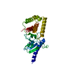
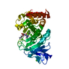
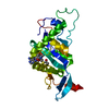
- PDB-6z2t: Three-dimensional structure of an influenza hemagglutinin LAH pro... -
+ Open data

- Basic information
| Entry | Database: PDB / ID: 6z2t | ||||||
|---|---|---|---|---|---|---|---|
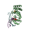
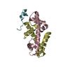
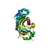
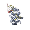
| Title | Three-dimensional structure of an influenza hemagglutinin LAH protein in its post-fusion conformation | ||||||
 Components Components | Hemagglutinin | ||||||
 Keywords Keywords | VIRAL PROTEIN / Hemagglutinin stem / Long alpha helix / Influenza / Post-fusion conformation / Tri-stalk protein | ||||||
| Function / homology | Haemagglutinin, influenzavirus B / Haemagglutinin / Haemagglutinin, influenzavirus A/B / host cell surface receptor binding / fusion of virus membrane with host plasma membrane / viral envelope / membrane / PHOSPHATE ION / Hemagglutinin Function and homology information Function and homology information | ||||||
| Biological species |   Influenza A virus Influenza A virus | ||||||
| Method |  X-RAY DIFFRACTION / SYNCHROTRON / MOLECULAR REPLACEMENT / Resolution: 1.34 Å X-RAY DIFFRACTION / SYNCHROTRON / MOLECULAR REPLACEMENT / Resolution: 1.34 Å | ||||||
 Authors Authors | Kirsteina, A. / Kazaks, A. / Tars, K. | ||||||
 Citation Citation | Journal: To Be Published Title: Three-dimensional structure of an influenza hemagglutinin LAH protein Authors: Kirsteina, A. / Kazaks, A. / Tars, K. | ||||||
| History |
|
- Structure visualization
| Structure viewer | Molecule: MolmilJmol/JSmol |
|---|
- Downloads & links
-Download
| PDBx/mmCIF format | 6z2t.cif.gz | 26.6 KB | Display | PDBx/mmCIF format |
|---|---|---|---|---|
| PDB format | pdb6z2t.ent.gz | 15.8 KB | Display | PDB format |
| PDBx/mmJSON format | 6z2t.json.gz | Tree view | PDBx/mmJSON format | |
| Others |  Other downloads Other downloads |
-Validation report
| Arichive directory | https://data.pdbj.org/pub/pdb/validation_reports/z2/6z2tftp://data.pdbj.org/pub/pdb/validation_reports/z2/6z2t | HTTPS FTP |
|---|
-Related structure data
| Related structure data |  6golS S: Starting model for refinement |
|---|---|
| Similar structure data |


-Links
 PDBj
PDBj


- Assembly
| Deposited unit | 
| ||||||||
|---|---|---|---|---|---|---|---|---|---|
| 1 | 
| ||||||||
| Unit cell |
| ||||||||
| Components on special symmetry positions |
|
-Components
| #1: Protein | Mass: 6964.825 Da / Num. of mol.: 1 Source method: isolated from a genetically manipulated source Source: (gene. exp.) Influenza A virus (A/Mexico City/63/2009(H1N1))Production host:  |
|---|---|
| #2: Chemical | ChemComp-PO4 /   Mass: 94.971 Da / Num. of mol.: 1 / Source method: obtained synthetically / Formula: PO4 Mass: 94.971 Da / Num. of mol.: 1 / Source method: obtained synthetically / Formula: PO4 |
| #3: Water | ChemComp-HOH /  Mass: 18.015 Da / Num. of mol.: 47 / Source method: isolated from a natural source / Formula: H2O Mass: 18.015 Da / Num. of mol.: 47 / Source method: isolated from a natural source / Formula: H2O |
| Has ligand of interest | N |
-Experimental details
-Experiment
| Experiment | Method: X-RAY DIFFRACTION / Number of used crystals: 1 |
|---|
- Sample preparation
| Crystal | Density Matthews: 2.32 Å3/Da / Density % sol: 47 % |
|---|---|
| Crystal grow | Temperature: 294 K / Method: vapor diffusion, sitting drop / pH: 6.7 Details: 0.15 M H6NO4P and 40% MPD; pH 6.7; protein 10 mg/mL |
-Data collection
| Diffraction | Mean temperature: 100 K / Serial crystal experiment: N |
|---|---|
| Diffraction source | Source: SYNCHROTRON / Site: MAX II  / Beamline: I911-3 / Wavelength: 0.91841 Å / Beamline: I911-3 / Wavelength: 0.91841 Å |
| Detector | Type: MARMOSAIC 225 mm CCD / Detector: CCD / Date: Apr 1, 2017 / Details: RH-COATED TOROIDAL SI MIRROR |
| Radiation | Monochromator: KMC-1 / Protocol: SINGLE WAVELENGTH / Monochromatic (M) / Laue (L): M / Scattering type: x-ray |
| Radiation wavelength | Wavelength: 0.91841 Å / Relative weight: 1 |
| Reflection | Resolution: 1.34→55.52 Å / Num. obs: 15916 / % possible obs: 99.5 % / Redundancy: 7.1 % / Biso Wilson estimate: 20.069 Å2 / Rmerge(I) obs: 0.05 / Net I/σ(I): 13.3 |
| Reflection shell | Resolution: 1.34→1.42 Å / Redundancy: 7 % / Rmerge(I) obs: 0.583 / Mean I/σ(I) obs: 2.5 / Num. unique obs: 2234 / % possible all: 98.4 |
- Processing
| Software |
| ||||||||||||||||||||||||||||||||||||||||||||||||||||||||||||
|---|---|---|---|---|---|---|---|---|---|---|---|---|---|---|---|---|---|---|---|---|---|---|---|---|---|---|---|---|---|---|---|---|---|---|---|---|---|---|---|---|---|---|---|---|---|---|---|---|---|---|---|---|---|---|---|---|---|---|---|---|---|
| Refinement | Method to determine structure: MOLECULAR REPLACEMENT Starting model: 6GOL Resolution: 1.34→41.68 Å / Cor.coef. Fo:Fc: 0.96 / Cor.coef. Fo:Fc free: 0.956 / SU B: 1.479 / SU ML: 0.055 / Cross valid method: THROUGHOUT / σ(F): 0 / ESU R: 0.06 / ESU R Free: 0.062 / Stereochemistry target values: MAXIMUM LIKELIHOOD Details: HYDROGENS HAVE BEEN ADDED IN THE RIDING POSITIONS U VALUES : REFINED INDIVIDUALLY
| ||||||||||||||||||||||||||||||||||||||||||||||||||||||||||||
| Solvent computation | Ion probe radii: 0.8 Å / Shrinkage radii: 0.8 Å / VDW probe radii: 1.2 Å / Solvent model: MASK | ||||||||||||||||||||||||||||||||||||||||||||||||||||||||||||
| Displacement parameters | Biso max: 76.34 Å2 / Biso mean: 25.219 Å2 / Biso min: 15.28 Å2
| ||||||||||||||||||||||||||||||||||||||||||||||||||||||||||||
| Refinement step | Cycle: final / Resolution: 1.34→41.68 Å
| ||||||||||||||||||||||||||||||||||||||||||||||||||||||||||||
| Refine LS restraints |
| ||||||||||||||||||||||||||||||||||||||||||||||||||||||||||||
| LS refinement shell | Resolution: 1.343→1.378 Å / Rfactor Rfree error: 0 / Total num. of bins used: 20
|
