 Movie
Movie Controller

[English] 日本語
 Yorodumi
- PDB-5imu: A fragment of conserved hypothetical protein Rv3899c (residues 18... -
+ Open data

- Basic information
| Entry | Database: PDB / ID: 5imu | ||||||
|---|---|---|---|---|---|---|---|
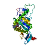
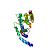
| Title | A fragment of conserved hypothetical protein Rv3899c (residues 184-410) from Mycobacterium tuberculosis | ||||||
 Components Components | Tat (Twin-arginine translocation) pathway signal sequence containing protein | ||||||
 Keywords Keywords | SIGNALING PROTEIN / alpha/beta/alpha sandwich / alpha helix bundule | ||||||
| Function / homology | Domain of unknown function DUF5632 / Protein of unknown function DUF5631 / Family of unknown function (DUF5631) / Family of unknown function (DUF5632) / extracellular region / : / Tat (Twin-arginine translocation) pathway signal sequence containing protein Function and homology information Function and homology information | ||||||
| Biological species |   Mycobacterium tuberculosis (bacteria) Mycobacterium tuberculosis (bacteria) | ||||||
| Method |  X-RAY DIFFRACTION / SIRAS / Resolution: 1.9 Å X-RAY DIFFRACTION / SIRAS / Resolution: 1.9 Å | ||||||
 Authors Authors | Li, D.F. / Gao, Y.R. / Liu, Y.Y. / Bi, L.J. | ||||||
 Citation Citation | Journal: Acta Crystallogr F Struct Biol Commun / Year: 2016 Title: Crystal structure of Rv3899c(184-410), a hypothetical protein from Mycobacterium tuberculosis Authors: Liu, Y.Y. / Gao, Y.R. / Li, D.F. / Fleming, J. / Li, H.L. / Bi, L.J. | ||||||
| History |
|
- Structure visualization
| Structure viewer | Molecule: MolmilJmol/JSmol |
|---|
- Downloads & links
-Download
| PDBx/mmCIF format | 5imu.cif.gz | 64 KB | Display | PDBx/mmCIF format |
|---|---|---|---|---|
| PDB format | pdb5imu.ent.gz | 45 KB | Display | PDB format |
| PDBx/mmJSON format | 5imu.json.gz | Tree view | PDBx/mmJSON format | |
| Others |  Other downloads Other downloads |
-Validation report
| Arichive directory | https://data.pdbj.org/pub/pdb/validation_reports/im/5imuftp://data.pdbj.org/pub/pdb/validation_reports/im/5imu | HTTPS FTP |
|---|
-Related structure data
| Similar structure data |
|---|


-Links
 PDBj
PDBj- Assembly
| Deposited unit | 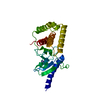
| ||||||||
|---|---|---|---|---|---|---|---|---|---|
| 1 |
| ||||||||
| Unit cell |
|
-Components
| #1: Protein | Mass: 24251.449 Da / Num. of mol.: 1 / Fragment: UNP residues 184-410 Source method: isolated from a genetically manipulated source Source: (gene. exp.) Mycobacterium tuberculosis (strain ATCC 25618 / H37Rv) (bacteria)Strain: ATCC 25618 / H37Rv / Gene: Rv3899c, LH57_21230 / Plasmid: pET-28a / Production host: |
|---|---|
| #2: Chemical | ChemComp-K /   Mass: 39.098 Da / Num. of mol.: 1 / Source method: obtained synthetically / Formula: K Mass: 39.098 Da / Num. of mol.: 1 / Source method: obtained synthetically / Formula: K |
| #3: Water | ChemComp-HOH /  Mass: 18.015 Da / Num. of mol.: 348 / Source method: isolated from a natural source / Formula: H2O Mass: 18.015 Da / Num. of mol.: 348 / Source method: isolated from a natural source / Formula: H2O |
-Experimental details
-Experiment
| Experiment | Method: X-RAY DIFFRACTION / Number of used crystals: 1 |
|---|
- Sample preparation
| Crystal | Density Matthews: 2.06 Å3/Da / Density % sol: 40.38 % |
|---|---|
| Crystal grow | Temperature: 289 K / Method: vapor diffusion, hanging drop Details: 0.2M ammonium acetate, 0.1M bis-tris (pH 5.5), 25% PEG 3350 PH range: 5.0-5.6 |
-Data collection
| Diffraction | Mean temperature: 100 K |
|---|---|
| Diffraction source | Source: ROTATING ANODE / Type: RIGAKU / Wavelength: 1.5418 Å |
| Detector | Type: RIGAKU RAXIS IV++ / Detector: IMAGE PLATE / Date: Feb 21, 2014 |
| Radiation | Protocol: SINGLE WAVELENGTH / Monochromatic (M) / Laue (L): M / Scattering type: x-ray |
| Radiation wavelength | Wavelength: 1.5418 Å / Relative weight: 1 |
| Reflection | Resolution: 1.9→44.51 Å / Num. obs: 16973 / % possible obs: 100 % / Redundancy: 6.6 % / Biso Wilson estimate: 17.4 Å2 / CC1/2: 0.997 / Rmerge(I) obs: 0.1 / Rsym value: 0.1 / Net I/σ(I): 13.1 |
| Reflection shell | Resolution: 1.9→2 Å / Redundancy: 4.3 % / Rmerge(I) obs: 0.425 / Mean I/σ(I) obs: 3.5 / % possible all: 93 |
- Processing
| Software |
| |||||||||||||||||||||||||||||||||||||||||||||||||
|---|---|---|---|---|---|---|---|---|---|---|---|---|---|---|---|---|---|---|---|---|---|---|---|---|---|---|---|---|---|---|---|---|---|---|---|---|---|---|---|---|---|---|---|---|---|---|---|---|---|---|
| Refinement | Method to determine structure: SIRAS / Resolution: 1.9→44.503 Å / SU ML: 0.19 / Cross valid method: FREE R-VALUE / σ(F): 1.36 / Phase error: 19
| |||||||||||||||||||||||||||||||||||||||||||||||||
| Solvent computation | Shrinkage radii: 0.9 Å / VDW probe radii: 1.11 Å | |||||||||||||||||||||||||||||||||||||||||||||||||
| Displacement parameters | Biso mean: 19.3 Å2 | |||||||||||||||||||||||||||||||||||||||||||||||||
| Refinement step | Cycle: LAST / Resolution: 1.9→44.503 Å
| |||||||||||||||||||||||||||||||||||||||||||||||||
| Refine LS restraints |
| |||||||||||||||||||||||||||||||||||||||||||||||||
| LS refinement shell |
|
