 Movie
Movie Controller

[English] 日本語
 Yorodumi
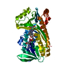
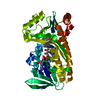
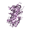
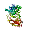
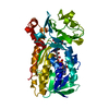
- PDB-6z2q: Crystal structure of wild type OgpA from Akkermansia muciniphila ... -
+ Open data

- Basic information
| Entry | Database: PDB / ID: 6z2q | ||||||
|---|---|---|---|---|---|---|---|
| Title | Crystal structure of wild type OgpA from Akkermansia muciniphila in complex with an O-glycopeptide (GalGalNAc-TS) product | ||||||
 Components Components |
| ||||||
 Keywords Keywords | HYDROLASE / O-glycan endopeptidase / mucins / OgpA. metalloprotease | ||||||
| Function / homology |  Function and homology information Function and homology informationpositive regulation of biosynthetic process of antibacterial peptides active against Gram-negative bacteria / defense response to insect / response to bacterium / defense response / antibacterial humoral response / defense response to Gram-negative bacterium / defense response to Gram-positive bacterium / defense response to bacterium / innate immune response / extracellular region / metal ion binding Similarity search - Function | ||||||
| Biological species |  Akkermansia muciniphila ATCC BAA-835 (bacteria) Akkermansia muciniphila ATCC BAA-835 (bacteria)synthetic construct (others) | ||||||
| Method |  X-RAY DIFFRACTION / SYNCHROTRON / MOLECULAR REPLACEMENT / Resolution: 2.347 Å X-RAY DIFFRACTION / SYNCHROTRON / MOLECULAR REPLACEMENT / Resolution: 2.347 Å | ||||||
 Authors Authors | Trastoy, B. / Naegali, A. / Anso, I. / Sjogren, J. / Guerin, M.E. | ||||||
 Citation Citation | Journal: Nat Commun / Year: 2020 Title: Structural basis of mammalian mucin processing by the human gut O-glycopeptidase OgpA from Akkermansia muciniphila. Authors: Trastoy, B. / Naegeli, A. / Anso, I. / Sjogren, J. / Guerin, M.E. | ||||||
| History |
|
- Structure visualization
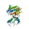
| Structure viewer | Molecule: MolmilJmol/JSmol |
|---|
- Downloads & links
-Download
| PDBx/mmCIF format | 6z2q.cif.gz | 91.3 KB | Display | PDBx/mmCIF format |
|---|---|---|---|---|
| PDB format | pdb6z2q.ent.gz | 65.2 KB | Display | PDB format |
| PDBx/mmJSON format | 6z2q.json.gz | Tree view | PDBx/mmJSON format | |
| Others |  Other downloads Other downloads |
-Validation report
| Summary document | 6z2q_validation.pdf.gz | 823.9 KB | Display | wwPDB validaton report |
|---|---|---|---|---|
| Full document | 6z2q_full_validation.pdf.gz | 827 KB | Display | |
| Data in XML | 6z2q_validation.xml.gz | 15.7 KB | Display | |
| Data in CIF | 6z2q_validation.cif.gz | 22.1 KB | Display | |
| Arichive directory | https://data.pdbj.org/pub/pdb/validation_reports/z2/6z2qftp://data.pdbj.org/pub/pdb/validation_reports/z2/6z2q | HTTPS FTP |
-Related structure data
| Related structure data |  6z2dSC 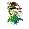 6z2oC  6z2pC S: Starting model for refinement C: citing same article ( |
|---|---|
| Similar structure data |

-Links
 PDBj
PDBj

- Assembly
| Deposited unit | 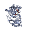
| ||||||||
|---|---|---|---|---|---|---|---|---|---|
| 1 |
| ||||||||
| Unit cell |
|
-Components
-Protein / Protein/peptide / Sugars , 3 types, 3 molecules AD
| #1: Protein | Mass: 41856.137 Da / Num. of mol.: 1 Source method: isolated from a genetically manipulated source Source: (gene. exp.) Akkermansia muciniphila ATCC BAA-835 (bacteria)Gene: Amuc_1119 / Production host: |
|---|---|
| #2: Protein/peptide | Mass: 2204.579 Da / Num. of mol.: 1 / Source method: obtained synthetically / Source: (synth.) synthetic construct (others) / References: UniProt: P36193*PLUS |
| #3: Polysaccharide | beta-D-galactopyranose-(1-3)-2-acetamido-2-deoxy-alpha-D-galactopyranose / Thomsen-Friedenreich antigen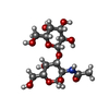  Source method: isolated from a genetically manipulated source Details: oligosaccharide / References: Thomsen-Friedenreich antigen |
-Non-polymers , 4 types, 99 molecules 


| #4: Chemical |  Mass: 65.409 Da / Num. of mol.: 2 Mass: 65.409 Da / Num. of mol.: 2Source method: isolated from a genetically manipulated source Formula: Zn #5: Chemical | ChemComp-ACY / |  Mass: 60.052 Da / Num. of mol.: 1 / Source method: obtained synthetically / Formula: C2H4O2 Mass: 60.052 Da / Num. of mol.: 1 / Source method: obtained synthetically / Formula: C2H4O2#6: Chemical |  Mass: 62.068 Da / Num. of mol.: 2 Mass: 62.068 Da / Num. of mol.: 2Source method: isolated from a genetically manipulated source Formula: C2H6O2 #7: Water | ChemComp-HOH / | Mass: 18.015 Da / Num. of mol.: 94 / Source method: isolated from a natural source / Formula: H2O |
|---|
-Details
| Has ligand of interest | Y |
|---|---|
| Has protein modification | Y |
-Experimental details
-Experiment
| Experiment | Method: X-RAY DIFFRACTION / Number of used crystals: 1 |
|---|
- Sample preparation
| Crystal | Density Matthews: 2.4 Å3/Da / Density % sol: 48.8 % |
|---|---|
| Crystal grow | Temperature: 291 K / Method: vapor diffusion, sitting drop Details: 100mM sodium HEPES/MOPS pH 7.5, 100mM carboxylic acids mixture (sodium formate, ammonium acetate, sodium citrate tribasic dihydrate, sodium potassium tartrate tetrahydrate and sodium oxamate) ...Details: 100mM sodium HEPES/MOPS pH 7.5, 100mM carboxylic acids mixture (sodium formate, ammonium acetate, sodium citrate tribasic dihydrate, sodium potassium tartrate tetrahydrate and sodium oxamate) and 50% (w/v) of precipitant mix based on 40% (w/v) PEG 500 MME and 20% (w/v) PEG 20,000 (Morpheus protein crystallization screen). |
-Data collection
| Diffraction | Mean temperature: 100 K / Serial crystal experiment: N |
|---|---|
| Diffraction source | Source: SYNCHROTRON / Site: Diamond  / Beamline: I03 / Wavelength: 0.97622 Å / Beamline: I03 / Wavelength: 0.97622 Å |
| Detector | Type: DECTRIS EIGER2 XE 16M / Detector: PIXEL / Date: Nov 27, 2019 |
| Radiation | Protocol: SINGLE WAVELENGTH / Monochromatic (M) / Laue (L): M / Scattering type: x-ray |
| Radiation wavelength | Wavelength: 0.97622 Å / Relative weight: 1 |
| Reflection | Resolution: 2.347→29.233 Å / Num. obs: 17627 / % possible obs: 98.62 % / Redundancy: 12.7 % / Biso Wilson estimate: 35.98 Å2 / CC1/2: 0.999 / CC star: 0.999 / Rmerge(I) obs: 0.1336 / Rrim(I) all: 0.1391 / Net I/σ(I): 19.29 |
| Reflection shell | Resolution: 2.347→2.43 Å / Redundancy: 10.6 % / Rmerge(I) obs: 0.96 / Mean I/σ(I) obs: 2.91 / Num. unique obs: 1530 / CC1/2: 0.2949 / CC star: 0.8 / Rrim(I) all: 1.007 / % possible all: 87.13 |
- Processing
| Software |
| ||||||||||||||||||||||||||||||||||||||||||||||||
|---|---|---|---|---|---|---|---|---|---|---|---|---|---|---|---|---|---|---|---|---|---|---|---|---|---|---|---|---|---|---|---|---|---|---|---|---|---|---|---|---|---|---|---|---|---|---|---|---|---|
| Refinement | Method to determine structure: MOLECULAR REPLACEMENT Starting model: 6Z2D Resolution: 2.347→29.233 Å / SU ML: 0.29 / Cross valid method: THROUGHOUT / σ(F): 1.34 / Phase error: 26.21 / Stereochemistry target values: ML
| ||||||||||||||||||||||||||||||||||||||||||||||||
| Solvent computation | Shrinkage radii: 0.9 Å / VDW probe radii: 1.11 Å / Solvent model: FLAT BULK SOLVENT MODEL | ||||||||||||||||||||||||||||||||||||||||||||||||
| Displacement parameters | Biso max: 74.02 Å2 / Biso mean: 37.1118 Å2 / Biso min: 25.72 Å2 | ||||||||||||||||||||||||||||||||||||||||||||||||
| Refinement step | Cycle: final / Resolution: 2.347→29.233 Å
| ||||||||||||||||||||||||||||||||||||||||||||||||
| LS refinement shell | Refine-ID: X-RAY DIFFRACTION / Rfactor Rfree error: 0
|
