 Movie
Movie Controller


+ Open data

- Basic information
| Entry | Database: PDB / ID: 6yu2 | |||||||||||||||
|---|---|---|---|---|---|---|---|---|---|---|---|---|---|---|---|---|
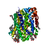
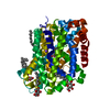
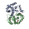
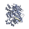
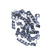
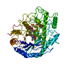
| Title | Crystal structure of MhsT in complex with L-isoleucine | |||||||||||||||
 Components Components | Sodium-dependent transporter | |||||||||||||||
 Keywords Keywords | TRANSPORT PROTEIN / LeuT-fold / amino acid transporter / L-isoleucine | |||||||||||||||
| Function / homology | : / Sodium:neurotransmitter symporter / Sodium:neurotransmitter symporter superfamily / Sodium:neurotransmitter symporter family / Sodium:neurotransmitter symporter family profile. / membrane / ISOLEUCINE / Na+-dependent transporter / Sodium-dependent transporter Function and homology information Function and homology information | |||||||||||||||
| Biological species |  Bacillus halodurans (bacteria) Bacillus halodurans (bacteria) | |||||||||||||||
| Method |  X-RAY DIFFRACTION / SYNCHROTRON / MOLECULAR REPLACEMENT / Resolution: 3.1 Å X-RAY DIFFRACTION / SYNCHROTRON / MOLECULAR REPLACEMENT / Resolution: 3.1 Å | |||||||||||||||
 Authors Authors | Focht, D. / Neumann, C. / Lyons, J. / Eguskiza Bilbao, A. / Blunck, R. / Malinauskaite, L. / Schwarz, I.O. / Javitch, J.A. / Quick, M. / Nissen, P. | |||||||||||||||
| Funding support |  Denmark, 4items Denmark, 4items
| |||||||||||||||
 Citation Citation | Journal: Embo J. / Year: 2021 Title: A non-helical region in transmembrane helix 6 of hydrophobic amino acid transporter MhsT mediates substrate recognition. Authors: Focht, D. / Neumann, C. / Lyons, J. / Eguskiza Bilbao, A. / Blunck, R. / Malinauskaite, L. / Schwarz, I.O. / Javitch, J.A. / Quick, M. / Nissen, P. | |||||||||||||||
| History |
|
- Structure visualization
| Structure viewer | Molecule: MolmilJmol/JSmol |
|---|
- Downloads & links
-Download
| PDBx/mmCIF format | 6yu2.cif.gz | 175.5 KB | Display | PDBx/mmCIF format |
|---|---|---|---|---|
| PDB format | pdb6yu2.ent.gz | 137.1 KB | Display | PDB format |
| PDBx/mmJSON format | 6yu2.json.gz | Tree view | PDBx/mmJSON format | |
| Others |  Other downloads Other downloads |
-Validation report
| Arichive directory | https://data.pdbj.org/pub/pdb/validation_reports/yu/6yu2ftp://data.pdbj.org/pub/pdb/validation_reports/yu/6yu2 | HTTPS FTP |
|---|
-Related structure data
| Related structure data |  6yu3C 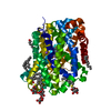 6yu4C 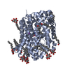 6yu5C  6yu6C 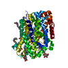 6yu7C  4us3S S: Starting model for refinement C: citing same article ( |
|---|---|
| Similar structure data |


-Links
 PDBj
PDBj


- Assembly
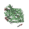
| Deposited unit | 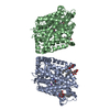
| ||||||||
|---|---|---|---|---|---|---|---|---|---|
| 1 | 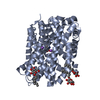
| ||||||||
| 2 | 
| ||||||||
| Unit cell |
|
-Components
| #1: Protein | Mass: 48360.844 Da / Num. of mol.: 2 Source method: isolated from a genetically manipulated source Source: (gene. exp.) Bacillus halodurans (bacteria) / Gene: E2L07_01100 / Production host: Lactococcus lactis (lactic acid bacteria) / References: UniProt: A0A4Y7X244, UniProt: Q9KDT3*PLUS#2: Sugar | ChemComp-BOG /   Type: D-saccharide / Mass: 292.369 Da / Num. of mol.: 6 Type: D-saccharide / Mass: 292.369 Da / Num. of mol.: 6Source method: isolated from a genetically manipulated source Formula: C14H28O6 / Comment: detergent*YM #3: Chemical | ChemComp-NA /   Mass: 22.990 Da / Num. of mol.: 4 / Source method: obtained synthetically / Formula: Na / Feature type: SUBJECT OF INVESTIGATION Mass: 22.990 Da / Num. of mol.: 4 / Source method: obtained synthetically / Formula: Na / Feature type: SUBJECT OF INVESTIGATION#4: Chemical |   Type: L-peptide linking / Mass: 131.173 Da / Num. of mol.: 2 / Source method: obtained synthetically / Formula: C6H13NO2 / Feature type: SUBJECT OF INVESTIGATION Type: L-peptide linking / Mass: 131.173 Da / Num. of mol.: 2 / Source method: obtained synthetically / Formula: C6H13NO2 / Feature type: SUBJECT OF INVESTIGATION#5: Water | ChemComp-HOH / |  Mass: 18.015 Da / Num. of mol.: 17 / Source method: isolated from a natural source / Formula: H2O Mass: 18.015 Da / Num. of mol.: 17 / Source method: isolated from a natural source / Formula: H2OHas ligand of interest | Y | |
|---|
-Experimental details
-Experiment
| Experiment | Method: X-RAY DIFFRACTION / Number of used crystals: 1 |
|---|
- Sample preparation
| Crystal | Density Matthews: 2.44 Å3/Da / Density % sol: 49.55 % |
|---|---|
| Crystal grow | Temperature: 293.15 K / Method: vapor diffusion, hanging drop / pH: 7 Details: Crystallized using HiLiDe with DOPC as added lipid. Crystals were obtained in 14-24% PEG400, 0.3-0.5M NaCl, 0.1 M Tris-HCl or HEPES-NaOH pH 7.0, 5% Trimethylamine N-oxide (TMANO), 5% or 10% glycerol. |
-Data collection
| Diffraction | Mean temperature: 100 K / Serial crystal experiment: N |
|---|---|
| Diffraction source | Source: SYNCHROTRON / Site: SLS  / Beamline: X06SA / Wavelength: 1.00001 Å / Beamline: X06SA / Wavelength: 1.00001 Å |
| Detector | Type: DECTRIS EIGER X 16M / Detector: PIXEL / Date: Apr 2, 2017 |
| Radiation | Protocol: SINGLE WAVELENGTH / Monochromatic (M) / Laue (L): M / Scattering type: x-ray |
| Radiation wavelength | Wavelength: 1.00001 Å / Relative weight: 1 |
| Reflection | Resolution: 3.1→43.7 Å / Num. obs: 13856 / % possible obs: 81.6 % / Redundancy: 2.7 % / CC1/2: 0.989 / Rmerge(I) obs: 0.195 / Net I/σ(I): 3.6 |
| Reflection shell | Resolution: 3.1→3.31 Å / Num. unique obs: 2557 / CC1/2: 0.636 |
- Processing
| Software |
| ||||||||||||||||||||||||||||||||||||
|---|---|---|---|---|---|---|---|---|---|---|---|---|---|---|---|---|---|---|---|---|---|---|---|---|---|---|---|---|---|---|---|---|---|---|---|---|---|
| Refinement | Method to determine structure: MOLECULAR REPLACEMENT Starting model: 4US3 Resolution: 3.1→43.698 Å / SU ML: 0.47 / Cross valid method: THROUGHOUT / σ(F): 1.35 / Phase error: 36.51
| ||||||||||||||||||||||||||||||||||||
| Solvent computation | Shrinkage radii: 0.9 Å / VDW probe radii: 1.11 Å | ||||||||||||||||||||||||||||||||||||
| Displacement parameters | Biso max: 73.23 Å2 / Biso mean: 56.685 Å2 / Biso min: 43.64 Å2 | ||||||||||||||||||||||||||||||||||||
| Refinement step | Cycle: final / Resolution: 3.1→43.698 Å
| ||||||||||||||||||||||||||||||||||||
| LS refinement shell | Refine-ID: X-RAY DIFFRACTION / Rfactor Rfree error: 0
|
