 Movie
Movie Controller

[English] 日本語
 Yorodumi
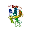
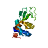
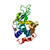
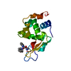
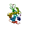
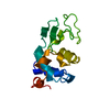
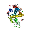
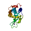
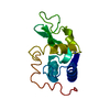
- PDB-6yod: Structure of Lysozyme from SiN IMISX setup collected by rotation ... -
+ Open data

- Basic information
| Entry | Database: PDB / ID: 6yod | ||||||
|---|---|---|---|---|---|---|---|
| Title | Structure of Lysozyme from SiN IMISX setup collected by rotation serial crystallography on crystals prelocated by 2D X-ray phase-contrast imaging | ||||||
 Components Components | Lysozyme | ||||||
 Keywords Keywords | HYDROLASE / 2D X-ray phase-contrast imaging / IMISX / in situ rotation images / prelocation / still images / serial crystallography | ||||||
| Function / homology |  Function and homology information Function and homology informationLactose synthesis / Antimicrobial peptides / Neutrophil degranulation / beta-N-acetylglucosaminidase activity / cell wall macromolecule catabolic process / lysozyme / lysozyme activity / killing of cells of another organism / defense response to Gram-negative bacterium / defense response to bacterium ...Lactose synthesis / Antimicrobial peptides / Neutrophil degranulation / beta-N-acetylglucosaminidase activity / cell wall macromolecule catabolic process / lysozyme / lysozyme activity / killing of cells of another organism / defense response to Gram-negative bacterium / defense response to bacterium / defense response to Gram-positive bacterium / endoplasmic reticulum / : / identical protein binding / cytoplasm Similarity search - Function | ||||||
| Biological species |  | ||||||
| Method |  X-RAY DIFFRACTION / SYNCHROTRON / MOLECULAR REPLACEMENT / Resolution: 1.601 Å X-RAY DIFFRACTION / SYNCHROTRON / MOLECULAR REPLACEMENT / Resolution: 1.601 Å | ||||||
 Authors Authors | Huang, C.-Y. / Martiel, I. / Villanueva-Perez, P. / Panepucci, E. / Caffrey, M. / Wang, M. | ||||||
 Citation Citation | Journal: Iucrj / Year: 2020 Title: Low-dose in situ prelocation of protein microcrystals by 2D X-ray phase-contrast imaging for serial crystallography. Authors: Martiel, I. / Huang, C.Y. / Villanueva-Perez, P. / Panepucci, E. / Basu, S. / Caffrey, M. / Pedrini, B. / Bunk, O. / Stampanoni, M. / Wang, M. | ||||||
| History |
|
- Structure visualization
| Structure viewer | Molecule: MolmilJmol/JSmol |
|---|
- Downloads & links
-Download
| PDBx/mmCIF format | 6yod.cif.gz | 43.8 KB | Display | PDBx/mmCIF format |
|---|---|---|---|---|
| PDB format | pdb6yod.ent.gz | 28.4 KB | Display | PDB format |
| PDBx/mmJSON format | 6yod.json.gz | Tree view | PDBx/mmJSON format | |
| Others |  Other downloads Other downloads |
-Validation report
| Arichive directory | https://data.pdbj.org/pub/pdb/validation_reports/yo/6yodftp://data.pdbj.org/pub/pdb/validation_reports/yo/6yod | HTTPS FTP |
|---|
-Related structure data
| Related structure data | 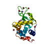 6yobC 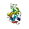 6yocC  6yoeC 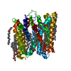 6yofC 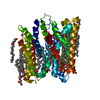 6yogC 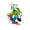 5d5fS S: Starting model for refinement C: citing same article ( |
|---|---|
| Similar structure data |

-Links
 PDBj
PDBj


- Assembly
| Deposited unit | 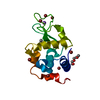
| ||||||||||||
|---|---|---|---|---|---|---|---|---|---|---|---|---|---|
| 1 |
| ||||||||||||
| Unit cell |
| ||||||||||||
| Components on special symmetry positions |
|
-Components
-Protein , 1 types, 1 molecules A
| #1: Protein | Mass: 14331.160 Da / Num. of mol.: 1 Source method: isolated from a genetically manipulated source Source: (gene. exp.) |
|---|
-Non-polymers , 6 types, 110 molecules 


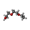

| #2: Chemical | ChemComp-BR /  Mass: 79.904 Da / Num. of mol.: 7 / Source method: obtained synthetically / Formula: Br Mass: 79.904 Da / Num. of mol.: 7 / Source method: obtained synthetically / Formula: Br#3: Chemical | ChemComp-NA / |  Mass: 22.990 Da / Num. of mol.: 1 / Source method: obtained synthetically / Formula: Na Mass: 22.990 Da / Num. of mol.: 1 / Source method: obtained synthetically / Formula: Na#4: Chemical | ChemComp-ACY / |  Mass: 60.052 Da / Num. of mol.: 1 / Source method: obtained synthetically / Formula: C2H4O2 Mass: 60.052 Da / Num. of mol.: 1 / Source method: obtained synthetically / Formula: C2H4O2#5: Chemical | ChemComp-PG0 / |  Mass: 120.147 Da / Num. of mol.: 1 / Source method: obtained synthetically / Formula: C5H12O3 / Comment: inhibitor, precipitant*YM Mass: 120.147 Da / Num. of mol.: 1 / Source method: obtained synthetically / Formula: C5H12O3 / Comment: inhibitor, precipitant*YM#6: Chemical | ChemComp-AE3 / |  Mass: 134.174 Da / Num. of mol.: 1 / Source method: obtained synthetically / Formula: C6H14O3 Mass: 134.174 Da / Num. of mol.: 1 / Source method: obtained synthetically / Formula: C6H14O3#7: Water | ChemComp-HOH / | Mass: 18.015 Da / Num. of mol.: 99 / Source method: isolated from a natural source / Formula: H2O |
|---|
-Details
| Has ligand of interest | N |
|---|---|
| Has protein modification | Y |
-Experimental details
-Experiment
| Experiment | Method: X-RAY DIFFRACTION / Number of used crystals: 1 |
|---|
- Sample preparation
| Crystal | Density Matthews: 2.08 Å3/Da / Density % sol: 40.82 % |
|---|---|
| Crystal grow | Temperature: 293 K / Method: lipidic cubic phase / pH: 4.5 Details: 0.5-1 M NaBr, 50-100 mM CH3COONa, pH 4.5, and 15-30 %(v/v) PEG400 |
-Data collection
| Diffraction | Mean temperature: 100 K / Serial crystal experiment: Y |
|---|---|
| Diffraction source | Source: SYNCHROTRON / Site: SLS  / Beamline: X06SA / Wavelength: 1 Å / Beamline: X06SA / Wavelength: 1 Å |
| Detector | Type: DECTRIS EIGER X 16M / Detector: PIXEL / Date: Sep 5, 2017 |
| Radiation | Protocol: SINGLE WAVELENGTH / Monochromatic (M) / Laue (L): M / Scattering type: x-ray |
| Radiation wavelength | Wavelength: 1 Å / Relative weight: 1 |
| Reflection | Resolution: 1.6→39.495 Å / Num. obs: 16419 / % possible obs: 99.87 % / Redundancy: 40.98 % / CC1/2: 0.99 / Rrim(I) all: 0.21 / Net I/σ(I): 13.58 |
| Reflection shell | Resolution: 1.6→1.64 Å / Redundancy: 36.73 % / Mean I/σ(I) obs: 0.86 / Num. unique obs: 1147 / CC1/2: 0.26 / Rrim(I) all: 12.17 |
| Serial crystallography sample delivery | Method: fixed target |
| Serial crystallography sample delivery fixed target | Description: IMISX / Sample dehydration prevention: SiN sandwich / Sample holding: SiN / Support base: 3D-printed holder in standard goniometer base |
- Processing
| Software |
| |||||||||||||||||||||||||||||||||||
|---|---|---|---|---|---|---|---|---|---|---|---|---|---|---|---|---|---|---|---|---|---|---|---|---|---|---|---|---|---|---|---|---|---|---|---|---|
| Refinement | Method to determine structure: MOLECULAR REPLACEMENT Starting model: 5D5F Resolution: 1.601→39.495 Å / SU ML: 0.17 / Cross valid method: THROUGHOUT / σ(F): 1.32 / Phase error: 23.5
| |||||||||||||||||||||||||||||||||||
| Solvent computation | Shrinkage radii: 0.9 Å / VDW probe radii: 1.11 Å | |||||||||||||||||||||||||||||||||||
| Displacement parameters | Biso max: 70.26 Å2 / Biso mean: 32.6264 Å2 / Biso min: 20.3 Å2 | |||||||||||||||||||||||||||||||||||
| Refinement step | Cycle: final / Resolution: 1.601→39.495 Å
| |||||||||||||||||||||||||||||||||||
| LS refinement shell | Refine-ID: X-RAY DIFFRACTION / Rfactor Rfree error: 0 / % reflection obs: 100 %
|
