 Movie
Movie Controller


+ Open data

- Basic information
| Entry | Database: PDB / ID: 6yex | ||||||
|---|---|---|---|---|---|---|---|
| Title | VcaM4I restriction endonuclease in the absence of DNA | ||||||
 Components Components | HNH endonuclease | ||||||
 Keywords Keywords | HYDROLASE / PUA superfamily / EVE domain / DNA endonuclease / modification-dependent restriction endonuclease / MDRE / single stranded DNA | ||||||
| Function / homology | HNH endonuclease / HNH nuclease / endonuclease activity / HNH endonuclease Function and homology information Function and homology information | ||||||
| Biological species |  Vibrio campbellii (bacteria) Vibrio campbellii (bacteria) | ||||||
| Method |  X-RAY DIFFRACTION / SYNCHROTRON / SAD / Resolution: 1.5 Å X-RAY DIFFRACTION / SYNCHROTRON / SAD / Resolution: 1.5 Å | ||||||
 Authors Authors | Pastor, M. / Czapinska, H. / Lutz, T. / Helbrecht, I. / Xu, S. / Bochtler, M. | ||||||
 Citation Citation | Journal: Nucleic Acids Res. / Year: 2021 Title: Crystal structures of the EVE-HNH endonuclease VcaM4I in the presence and absence of DNA. Authors: Pastor, M. / Czapinska, H. / Helbrecht, I. / Krakowska, K. / Lutz, T. / Xu, S.Y. / Bochtler, M. #1: Journal: Nucleic Acids Res. / Year: 2019 Title: A protein architecture guided screen for modification dependent restriction endonucleases. Authors: Lutz, T. / Flodman, K. / Copelas, A. / Czapinska, H. / Mabuchi, M. / Fomenkov, A. / He, X. / Bochtler, M. / Xu, S.Y. | ||||||
| History |
|
- Structure visualization
| Structure viewer | Molecule: MolmilJmol/JSmol |
|---|
- Downloads & links
-Download
| PDBx/mmCIF format | 6yex.cif.gz | 324.8 KB | Display | PDBx/mmCIF format |
|---|---|---|---|---|
| PDB format | pdb6yex.ent.gz | 266.1 KB | Display | PDB format |
| PDBx/mmJSON format | 6yex.json.gz | Tree view | PDBx/mmJSON format | |
| Others |  Other downloads Other downloads |
-Validation report
| Arichive directory | https://data.pdbj.org/pub/pdb/validation_reports/ye/6yexftp://data.pdbj.org/pub/pdb/validation_reports/ye/6yex | HTTPS FTP |
|---|
-Related structure data


-Links
 PDBj
PDBj
- Assembly
| Deposited unit | 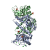
| ||||||||
|---|---|---|---|---|---|---|---|---|---|
| 1 |
| ||||||||
| Unit cell |
| ||||||||
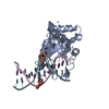
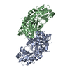
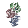
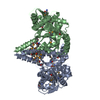
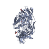
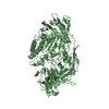
| Details | The modification specific VcaM4I restriction endonuclease is active as a dimer (each protomer cleaves one strand of the double stranded DNA substrate). The C-terminal HNH nuclease domains mediate the dimerization. The N-terminal EVE domains are responsible for the modification specificity. |
-Components
| #1: Protein | Mass: 35390.543 Da / Num. of mol.: 2 Source method: isolated from a genetically manipulated source Source: (gene. exp.) Vibrio campbellii (bacteria) / Gene: DSB67_20905 / Plasmid: pTXB1 (modified) / Production host: #2: Chemical | ChemComp-SO4 /   Mass: 96.063 Da / Num. of mol.: 7 / Source method: obtained synthetically / Formula: SO4 Mass: 96.063 Da / Num. of mol.: 7 / Source method: obtained synthetically / Formula: SO4#3: Chemical | ChemComp-CL /   Mass: 35.453 Da / Num. of mol.: 12 / Source method: isolated from a natural source / Formula: Cl Mass: 35.453 Da / Num. of mol.: 12 / Source method: isolated from a natural source / Formula: Cl#4: Water | ChemComp-HOH / |  Mass: 18.015 Da / Num. of mol.: 1024 / Source method: isolated from a natural source / Formula: H2O Mass: 18.015 Da / Num. of mol.: 1024 / Source method: isolated from a natural source / Formula: H2OHas ligand of interest | N | |
|---|
-Experimental details
-Experiment
| Experiment | Method: X-RAY DIFFRACTION / Number of used crystals: 1 |
|---|
- Sample preparation
| Crystal | Density Matthews: 2.38 Å3/Da / Density % sol: 48.24 % |
|---|---|
| Crystal grow | Temperature: 291 K / Method: vapor diffusion, hanging drop / pH: 8.5 Details: Protein (7.2 mg/ml) in 0.27 M NaCl, 13.5 mM Tris-HCl pH 8.5, 0.2 M sodium malonate and 0.9 mM TCEP was mixed in 1:1 ratio with reservoir solution (1.8 M ammonium sulfate, 0.1 M MES pH 5.25). ...Details: Protein (7.2 mg/ml) in 0.27 M NaCl, 13.5 mM Tris-HCl pH 8.5, 0.2 M sodium malonate and 0.9 mM TCEP was mixed in 1:1 ratio with reservoir solution (1.8 M ammonium sulfate, 0.1 M MES pH 5.25). Perfluoropolyether oil was used as a cryoprotectant. |
-Data collection
| Diffraction | Mean temperature: 100 K / Serial crystal experiment: N |
|---|---|
| Diffraction source | Source: SYNCHROTRON / Site: EMBL/DESY, HAMBURG  / Beamline: X13 / Wavelength: 1.0064 Å / Beamline: X13 / Wavelength: 1.0064 Å |
| Detector | Type: DECTRIS PILATUS 6M-F / Detector: PIXEL / Date: Oct 5, 2019 |
| Radiation | Protocol: SINGLE WAVELENGTH / Monochromatic (M) / Laue (L): M / Scattering type: x-ray |
| Radiation wavelength | Wavelength: 1.0064 Å / Relative weight: 1 |
| Reflection | Resolution: 1.5→30 Å / Num. obs: 107966 / % possible obs: 99.2 % / Redundancy: 13.1 % / Biso Wilson estimate: 32.7 Å2 / CC1/2: 1 / Rrim(I) all: 0.046 / Rsym value: 0.044 / Net I/σ(I): 26.06 |
| Reflection shell | Resolution: 1.5→1.59 Å / Redundancy: 13.4 % / Mean I/σ(I) obs: 2.27 / Num. unique obs: 17067 / CC1/2: 0.851 / Rrim(I) all: 1.104 / Rsym value: 1.062 / % possible all: 98.2 |
- Processing
| Software |
| |||||||||||||||||||||||||||||||||||||||||||||||||||||||||||||||||||||||||||||||||||||||||||||||||||||||||||||||||||||||||||||
|---|---|---|---|---|---|---|---|---|---|---|---|---|---|---|---|---|---|---|---|---|---|---|---|---|---|---|---|---|---|---|---|---|---|---|---|---|---|---|---|---|---|---|---|---|---|---|---|---|---|---|---|---|---|---|---|---|---|---|---|---|---|---|---|---|---|---|---|---|---|---|---|---|---|---|---|---|---|---|---|---|---|---|---|---|---|---|---|---|---|---|---|---|---|---|---|---|---|---|---|---|---|---|---|---|---|---|---|---|---|---|---|---|---|---|---|---|---|---|---|---|---|---|---|---|---|---|
| Refinement | Method to determine structure: SAD / Resolution: 1.5→28.48 Å / Cor.coef. Fo:Fc: 0.97 / Cor.coef. Fo:Fc free: 0.965 / SU B: 5.305 / SU ML: 0.082 / Cross valid method: THROUGHOUT / σ(F): 0 / ESU R: 0.083 / ESU R Free: 0.08 Details: HYDROGENS HAVE BEEN ADDED IN THE RIDING POSITIONS U VALUES : WITH TLS ADDED THE IDENTITY OF THE SOLVENT MOLECULES HAS BEEN ASSIGNED TENTATIVELY.
| |||||||||||||||||||||||||||||||||||||||||||||||||||||||||||||||||||||||||||||||||||||||||||||||||||||||||||||||||||||||||||||
| Solvent computation | Ion probe radii: 0.8 Å / Shrinkage radii: 0.8 Å / VDW probe radii: 1.2 Å | |||||||||||||||||||||||||||||||||||||||||||||||||||||||||||||||||||||||||||||||||||||||||||||||||||||||||||||||||||||||||||||
| Displacement parameters | Biso max: 109.92 Å2 / Biso mean: 33.633 Å2 / Biso min: 18.07 Å2
| |||||||||||||||||||||||||||||||||||||||||||||||||||||||||||||||||||||||||||||||||||||||||||||||||||||||||||||||||||||||||||||
| Refinement step | Cycle: final / Resolution: 1.5→28.48 Å
| |||||||||||||||||||||||||||||||||||||||||||||||||||||||||||||||||||||||||||||||||||||||||||||||||||||||||||||||||||||||||||||
| Refine LS restraints |
| |||||||||||||||||||||||||||||||||||||||||||||||||||||||||||||||||||||||||||||||||||||||||||||||||||||||||||||||||||||||||||||
| LS refinement shell | Resolution: 1.5→1.58 Å / Rfactor Rfree error: 0
| |||||||||||||||||||||||||||||||||||||||||||||||||||||||||||||||||||||||||||||||||||||||||||||||||||||||||||||||||||||||||||||
| Refinement TLS params. | Method: refined / Refine-ID: X-RAY DIFFRACTION
| |||||||||||||||||||||||||||||||||||||||||||||||||||||||||||||||||||||||||||||||||||||||||||||||||||||||||||||||||||||||||||||
| Refinement TLS group |
|
