 Movie
Movie Controller

[English] 日本語
 Yorodumi
- PDB-6y8p: Crystal structure of SNAP-tag labeled with a benzyl-tetramethylrh... -
+ Open data

- Basic information
| Entry | Database: PDB / ID: 6y8p | ||||||
|---|---|---|---|---|---|---|---|
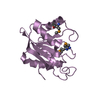
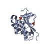
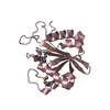
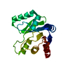
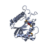
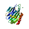
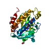
| Title | Crystal structure of SNAP-tag labeled with a benzyl-tetramethylrhodamine fluorophore | ||||||
 Components Components | O6-alkylguanine-DNA alkyltransferase mutant | ||||||
 Keywords Keywords | TRANSFERASE / SNAP-tag / self-labeling protein / tetramethylrhodamine / synthetic fluorophore | ||||||
| Function / homology |  Function and homology information Function and homology informationmethylated-DNA-[protein]-cysteine S-methyltransferase / methylated-DNA-[protein]-cysteine S-methyltransferase activity / methylation / DNA repair / DNA binding / metal ion binding / nucleus Similarity search - Function | ||||||
| Biological species |  Homo sapiens (human) Homo sapiens (human) | ||||||
| Method |  X-RAY DIFFRACTION / SYNCHROTRON / MOLECULAR REPLACEMENT / Resolution: 2.3 Å X-RAY DIFFRACTION / SYNCHROTRON / MOLECULAR REPLACEMENT / Resolution: 2.3 Å | ||||||
 Authors Authors | Gotthard, G. / Tanzer, T. / Johnsson, K. / Hiblot, J. | ||||||
 Citation Citation | Journal: Biochemistry / Year: 2021 Title: Kinetic and Structural Characterization of the Self-Labeling Protein Tags HaloTag7, SNAP-tag, and CLIP-tag. Authors: Wilhelm, J. / Kuhn, S. / Tarnawski, M. / Gotthard, G. / Tunnermann, J. / Tanzer, T. / Karpenko, J. / Mertes, N. / Xue, L. / Uhrig, U. / Reinstein, J. / Hiblot, J. / Johnsson, K. #1: Journal: Biorxiv / Year: 2021Title: Kinetic and structural characterization of the self-labeling protein tags HaloTag7, SNAP-tag and CLIP-tag Authors: Wilhelm, J. / Kuhn, S. / Tarnawski, M. / Gotthard, G. / Tunnermann, J. / Tanzer, T. / Karpenko, J. / Mertes, N. / Xue, L. / Uhrig, U. / Reinstein, J. / Hiblot, J. / Johnsson, K. | ||||||
| History |
|
- Structure visualization
| Structure viewer | Molecule: MolmilJmol/JSmol |
|---|
- Downloads & links
-Download
| PDBx/mmCIF format | 6y8p.cif.gz | 85.9 KB | Display | PDBx/mmCIF format |
|---|---|---|---|---|
| PDB format | pdb6y8p.ent.gz | 62.8 KB | Display | PDB format |
| PDBx/mmJSON format | 6y8p.json.gz | Tree view | PDBx/mmJSON format | |
| Others |  Other downloads Other downloads |
-Validation report
| Arichive directory | https://data.pdbj.org/pub/pdb/validation_reports/y8/6y8pftp://data.pdbj.org/pub/pdb/validation_reports/y8/6y8p | HTTPS FTP |
|---|
-Related structure data
| Related structure data | 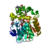 6y7aC 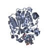 6y7bC 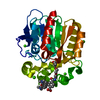 6zccC  3l00S S: Starting model for refinement C: citing same article ( |
|---|---|
| Similar structure data |


-Links
 PDBj
PDBj- Assembly
| Deposited unit | 
| ||||||||
|---|---|---|---|---|---|---|---|---|---|
| 1 |
| ||||||||
| Unit cell |
|
-Components
| #1: Protein | Mass: 18951.752 Da / Num. of mol.: 1 Source method: isolated from a genetically manipulated source Source: (gene. exp.) Homo sapiens (human) / Plasmid: pET51b / Production host:  |
|---|---|
| #2: Chemical | ChemComp-ZN /   Mass: 65.409 Da / Num. of mol.: 1 / Source method: obtained synthetically / Formula: Zn Mass: 65.409 Da / Num. of mol.: 1 / Source method: obtained synthetically / Formula: Zn |
| #3: Chemical | ChemComp-OGQ / [  Mass: 534.625 Da / Num. of mol.: 1 / Source method: obtained synthetically / Formula: C33H32N3O4 / Feature type: SUBJECT OF INVESTIGATION Mass: 534.625 Da / Num. of mol.: 1 / Source method: obtained synthetically / Formula: C33H32N3O4 / Feature type: SUBJECT OF INVESTIGATION |
| #4: Chemical | ChemComp-EDO /   Mass: 62.068 Da / Num. of mol.: 1 / Source method: obtained synthetically / Formula: C2H6O2 Mass: 62.068 Da / Num. of mol.: 1 / Source method: obtained synthetically / Formula: C2H6O2 |
| #5: Water | ChemComp-HOH /  Mass: 18.015 Da / Num. of mol.: 50 / Source method: isolated from a natural source / Formula: H2O Mass: 18.015 Da / Num. of mol.: 50 / Source method: isolated from a natural source / Formula: H2O |
| Has ligand of interest | Y |
| Has protein modification | Y |
-Experimental details
-Experiment
| Experiment | Method: X-RAY DIFFRACTION / Number of used crystals: 1 |
|---|
- Sample preparation
| Crystal | Density Matthews: 3.48 Å3/Da / Density % sol: 61.24 % / Description: Red hexaedral crystals |
|---|---|
| Crystal grow | Temperature: 291 K / Method: vapor diffusion, sitting drop / pH: 7.5 Details: 100 mM Na HEPES pH 7.5 25% PEG 8000 10 mg/mL protein 100 nl:100 nL |
-Data collection
| Diffraction | Mean temperature: 100 K / Serial crystal experiment: N |
|---|---|
| Diffraction source | Source: SYNCHROTRON / Site: ESRF  / Beamline: ID29 / Wavelength: 0.976 Å / Beamline: ID29 / Wavelength: 0.976 Å |
| Detector | Type: DECTRIS PILATUS3 S 6M / Detector: PIXEL / Date: Nov 3, 2016 |
| Radiation | Protocol: SINGLE WAVELENGTH / Monochromatic (M) / Laue (L): M / Scattering type: x-ray |
| Radiation wavelength | Wavelength: 0.976 Å / Relative weight: 1 |
| Reflection | Resolution: 2.3→48.98 Å / Num. obs: 11154 / % possible obs: 99.94 % / Redundancy: 10.7 % / CC1/2: 0.999 / Net I/σ(I): 15 |
| Reflection shell | Resolution: 2.3→2.382 Å / Num. unique obs: 1087 / CC1/2: 0.193 |
- Processing
| Software |
| ||||||||||||||||||||||||||||||||||||||||
|---|---|---|---|---|---|---|---|---|---|---|---|---|---|---|---|---|---|---|---|---|---|---|---|---|---|---|---|---|---|---|---|---|---|---|---|---|---|---|---|---|---|
| Refinement | Method to determine structure: MOLECULAR REPLACEMENT Starting model: 3l00 Resolution: 2.3→48.98 Å / SU ML: 0.43 / Cross valid method: THROUGHOUT / σ(F): 1.38 / Phase error: 37.4
| ||||||||||||||||||||||||||||||||||||||||
| Solvent computation | Shrinkage radii: 0.9 Å / VDW probe radii: 1.11 Å | ||||||||||||||||||||||||||||||||||||||||
| Displacement parameters | Biso max: 143.77 Å2 / Biso mean: 73.1096 Å2 / Biso min: 31.06 Å2 | ||||||||||||||||||||||||||||||||||||||||
| Refinement step | Cycle: final / Resolution: 2.3→48.98 Å
| ||||||||||||||||||||||||||||||||||||||||
| LS refinement shell | Refine-ID: X-RAY DIFFRACTION / Rfactor Rfree error: 0 / Total num. of bins used: 4 / % reflection obs: 100 %
| ||||||||||||||||||||||||||||||||||||||||
| Refinement TLS params. | Method: refined / Origin x: 16.4644 Å / Origin y: -5.4937 Å / Origin z: 2.3287 Å
| ||||||||||||||||||||||||||||||||||||||||
| Refinement TLS group |
|
