 Movie
Movie Controller


+ Open data

- Basic information
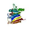
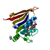
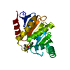
| Entry | Database: PDB / ID: 6y7g | ||||||
|---|---|---|---|---|---|---|---|
| Title | Structure of the human RAB3C in complex with GDP | ||||||
 Components Components | Ras-related protein Rab-3C | ||||||
 Keywords Keywords | STRUCTURAL GENOMICS / RAB3C / human Ras-related protein Rab-3C / Structural Genomics Consortium / SGC | ||||||
| Function / homology |  Function and homology information Function and homology informationregulation of exocytosis / myosin V binding / RAB geranylgeranylation / GTP-dependent protein binding / antigen processing and presentation / exocytosis / protein secretion / synaptic vesicle / synaptic vesicle membrane / vesicle ...regulation of exocytosis / myosin V binding / RAB geranylgeranylation / GTP-dependent protein binding / antigen processing and presentation / exocytosis / protein secretion / synaptic vesicle / synaptic vesicle membrane / vesicle / endosome / GTPase activity / GTP binding / perinuclear region of cytoplasm / plasma membrane Similarity search - Function | ||||||
| Biological species |  Homo sapiens (human) Homo sapiens (human) | ||||||
| Method |  X-RAY DIFFRACTION / SYNCHROTRON / MOLECULAR REPLACEMENT / molecular replacement / Resolution: 2.3 Å X-RAY DIFFRACTION / SYNCHROTRON / MOLECULAR REPLACEMENT / molecular replacement / Resolution: 2.3 Å | ||||||
 Authors Authors | Diaz-Saez, L. / Jung, S. / Raux, B. / Burgess-Brown, N.A. / von Delft, F. / Arrowsmith, C.H. / Edwards, A. / Bountra, C. / Huber, K.V.M. / Structural Genomics Consortium (SGC) | ||||||
| Funding support | 1items
| ||||||
 Citation Citation | Journal: To Be Published Title: Structure of the human RAB3C in complex with GDP Authors: Diaz-Saez, L. / Jung, S. / Burgess-Brown, N.A. / von Delft, F. / Arrowsmith, C.H. / Edwards, A. / Bountra, C. / Huber, K.V.M. | ||||||
| History |
|
- Structure visualization
| Structure viewer | Molecule: MolmilJmol/JSmol |
|---|
- Downloads & links
-Download
| PDBx/mmCIF format | 6y7g.cif.gz | 88.8 KB | Display | PDBx/mmCIF format |
|---|---|---|---|---|
| PDB format | pdb6y7g.ent.gz | 64.6 KB | Display | PDB format |
| PDBx/mmJSON format | 6y7g.json.gz | Tree view | PDBx/mmJSON format | |
| Others |  Other downloads Other downloads |
-Validation report
| Summary document | 6y7g_validation.pdf.gz | 1.1 MB | Display | wwPDB validaton report |
|---|---|---|---|---|
| Full document | 6y7g_full_validation.pdf.gz | 1.1 MB | Display | |
| Data in XML | 6y7g_validation.xml.gz | 16 KB | Display | |
| Data in CIF | 6y7g_validation.cif.gz | 21.2 KB | Display | |
| Arichive directory | https://data.pdbj.org/pub/pdb/validation_reports/y7/6y7gftp://data.pdbj.org/pub/pdb/validation_reports/y7/6y7g | HTTPS FTP |
-Related structure data
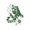
| Related structure data |  1zbdS S: Starting model for refinement |
|---|---|
| Similar structure data |


-Links
 PDBj
PDBj- Assembly
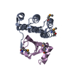
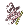
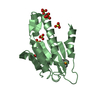
| Deposited unit | 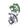
| ||||||||
|---|---|---|---|---|---|---|---|---|---|
| 1 | 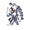
| ||||||||
| 2 | 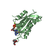
| ||||||||
| Unit cell |
|
-Components
| #1: Protein | Mass: 25980.168 Da / Num. of mol.: 2 Source method: isolated from a genetically manipulated source Source: (gene. exp.) Homo sapiens (human) / Gene: RAB3C / Plasmid: pET28a-LIC / Production host:  #2: Chemical |   Type: RNA linking / Mass: 443.201 Da / Num. of mol.: 2 / Source method: obtained synthetically / Formula: C10H15N5O11P2 / Feature type: SUBJECT OF INVESTIGATION / Comment: GDP, energy-carrying molecule*YM Type: RNA linking / Mass: 443.201 Da / Num. of mol.: 2 / Source method: obtained synthetically / Formula: C10H15N5O11P2 / Feature type: SUBJECT OF INVESTIGATION / Comment: GDP, energy-carrying molecule*YM#3: Chemical |   Mass: 209.240 Da / Num. of mol.: 2 / Source method: obtained synthetically / Formula: C8H19NO5 / Comment: pH buffer*YM Mass: 209.240 Da / Num. of mol.: 2 / Source method: obtained synthetically / Formula: C8H19NO5 / Comment: pH buffer*YM#4: Chemical |   Mass: 24.305 Da / Num. of mol.: 2 / Source method: obtained synthetically / Formula: Mg Mass: 24.305 Da / Num. of mol.: 2 / Source method: obtained synthetically / Formula: Mg#5: Water | ChemComp-HOH / |  Mass: 18.015 Da / Num. of mol.: 82 / Source method: isolated from a natural source / Formula: H2O Mass: 18.015 Da / Num. of mol.: 82 / Source method: isolated from a natural source / Formula: H2OHas ligand of interest | Y | |
|---|
-Experimental details
-Experiment
| Experiment | Method: X-RAY DIFFRACTION / Number of used crystals: 1 |
|---|
- Sample preparation
| Crystal grow | Temperature: 277 K / Method: vapor diffusion, sitting drop / pH: 6.5 / Details: 25% PEG3350, 0.1M BIS-TRIS pH 6.5 |
|---|
-Data collection
| Diffraction | Mean temperature: 80 K / Serial crystal experiment: N | ||||||||||||||||||||||||||||||
|---|---|---|---|---|---|---|---|---|---|---|---|---|---|---|---|---|---|---|---|---|---|---|---|---|---|---|---|---|---|---|---|
| Diffraction source | Source: SYNCHROTRON / Site: Diamond  / Beamline: I04 / Wavelength: 0.9795 Å / Beamline: I04 / Wavelength: 0.9795 Å | ||||||||||||||||||||||||||||||
| Detector | Type: DECTRIS EIGER2 XE 16M / Detector: PIXEL / Date: Dec 15, 2019 | ||||||||||||||||||||||||||||||
| Radiation | Protocol: SINGLE WAVELENGTH / Monochromatic (M) / Laue (L): M / Scattering type: x-ray | ||||||||||||||||||||||||||||||
| Radiation wavelength | Wavelength: 0.9795 Å / Relative weight: 1 | ||||||||||||||||||||||||||||||
| Reflection | Resolution: 2.3→63.78 Å / Num. obs: 16096 / % possible obs: 98.4 % / Redundancy: 3.6 % / CC1/2: 0.997 / Rmerge(I) obs: 0.08 / Rpim(I) all: 0.049 / Rrim(I) all: 0.094 / Net I/σ(I): 9.5 / Num. measured all: 57285 / Scaling rejects: 30 | ||||||||||||||||||||||||||||||
| Reflection shell | Diffraction-ID: 1
|
-Phasing
| Phasing | Method: molecular replacement | |||||||||
|---|---|---|---|---|---|---|---|---|---|---|
| Phasing MR | Model details: Phaser MODE: MR_AUTO
|
- Processing
| Software |
| ||||||||||||||||||||||||||||||||||||||||||||||||||||||||||||
|---|---|---|---|---|---|---|---|---|---|---|---|---|---|---|---|---|---|---|---|---|---|---|---|---|---|---|---|---|---|---|---|---|---|---|---|---|---|---|---|---|---|---|---|---|---|---|---|---|---|---|---|---|---|---|---|---|---|---|---|---|---|
| Refinement | Method to determine structure: MOLECULAR REPLACEMENT Starting model: 1ZBD Resolution: 2.3→63.78 Å / Cor.coef. Fo:Fc: 0.948 / Cor.coef. Fo:Fc free: 0.867 / SU B: 10.34 / SU ML: 0.242 / Cross valid method: THROUGHOUT / σ(F): 0 / ESU R: 0.395 / ESU R Free: 0.293 / Stereochemistry target values: MAXIMUM LIKELIHOOD Details: HYDROGENS HAVE BEEN ADDED IN THE RIDING POSITIONS U VALUES : REFINED INDIVIDUALLY
| ||||||||||||||||||||||||||||||||||||||||||||||||||||||||||||
| Solvent computation | Ion probe radii: 0.8 Å / Shrinkage radii: 0.8 Å / VDW probe radii: 1.2 Å / Solvent model: MASK | ||||||||||||||||||||||||||||||||||||||||||||||||||||||||||||
| Displacement parameters | Biso max: 99.42 Å2 / Biso mean: 43.127 Å2 / Biso min: 27.52 Å2
| ||||||||||||||||||||||||||||||||||||||||||||||||||||||||||||
| Refinement step | Cycle: final / Resolution: 2.3→63.78 Å
| ||||||||||||||||||||||||||||||||||||||||||||||||||||||||||||
| Refine LS restraints |
| ||||||||||||||||||||||||||||||||||||||||||||||||||||||||||||
| LS refinement shell | Resolution: 2.3→2.36 Å / Rfactor Rfree error: 0 / Total num. of bins used: 20
|
