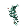
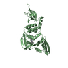
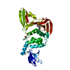
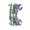
[English] 日本語
Yorodumi
- PDB-1zbd: STRUCTURAL BASIS OF RAB EFFECTOR SPECIFICITY: CRYSTAL STRUCTURE O... -

+
Open data


ID or keywords:

Loading...

-
Basic information

Entry
Database: PDB / ID: 1zbd
TitleSTRUCTURAL BASIS OF RAB EFFECTOR SPECIFICITY: CRYSTAL STRUCTURE OF THE SMALL G PROTEIN RAB3A COMPLEXED WITH THE EFFECTOR DOMAIN OF RABPHILIN-3A
Components(RABPHILIN-3A) x 2
KeywordsG PROTEIN / EFFECTOR / RABCDR / SYNAPTIC EXOCYTOSIS / RAB PROTEIN / RAB3A / RABPHILIN
Function / homology
Function and homology information


GDP-dissociation inhibitor binding / evoked neurotransmitter secretion / acrosomal vesicle exocytosis / regulation of presynaptic dense core granule exocytosis / RAB GEFs exchange GTP for GDP on RABs / positive regulation of regulated secretory pathway / regulation of plasma membrane repair / maintenance of presynaptic active zone structure / regulation of synaptic vesicle fusion to presynaptic active zone membrane / sensory perception of touch ...GDP-dissociation inhibitor binding / evoked neurotransmitter secretion / acrosomal vesicle exocytosis / regulation of presynaptic dense core granule exocytosis / RAB GEFs exchange GTP for GDP on RABs / positive regulation of regulated secretory pathway / regulation of plasma membrane repair / maintenance of presynaptic active zone structure / regulation of synaptic vesicle fusion to presynaptic active zone membrane / sensory perception of touch / RAB geranylgeranylation / selenium binding / Rab protein signal transduction / synaptic vesicle recycling / spontaneous neurotransmitter secretion / respiratory system process / regulation of synaptic vesicle priming / Glutamate Neurotransmitter Release Cycle / Norepinephrine Neurotransmitter Release Cycle / Acetylcholine Neurotransmitter Release Cycle / Serotonin Neurotransmitter Release Cycle / GABA synthesis, release, reuptake and degradation / regulated exocytosis / Dopamine Neurotransmitter Release Cycle / extrinsic component of synaptic vesicle membrane / synaptic vesicle clustering / synaptic vesicle maturation / neuromuscular synaptic transmission / regulation of calcium ion-dependent exocytosis / cholinergic synapse / GTP-dependent protein binding / calcium-ion regulated exocytosis / myosin V binding / lysosome localization / plasma membrane repair / vesicle docking involved in exocytosis / regulation of exocytosis / inositol 1,4,5 trisphosphate binding / regulation of short-term neuronal synaptic plasticity / calcium-dependent phospholipid binding / dendritic spine organization / insulin secretion / synaptic vesicle priming / Neutrophil degranulation / synaptic vesicle transport / regulation of synaptic vesicle exocytosis / extrinsic component of membrane / presynaptic active zone / regulation of dopamine secretion / phosphate ion binding / exocytosis / synaptic vesicle exocytosis / ATPase activator activity / regulation of NMDA receptor activity / protein secretion / response to electrical stimulus / phosphatidylinositol-4,5-bisphosphate binding / mitochondrion organization / axonogenesis / post-embryonic development / acrosomal vesicle / secretory granule / establishment of localization in cell / protein localization to plasma membrane / phospholipid binding / lung development / intracellular protein transport / neuromuscular junction / terminal bouton / synaptic vesicle membrane / small GTPase binding / synaptic vesicle / protein-macromolecule adaptor activity / presynapse / ATPase binding / postsynaptic membrane / postsynapse / dendritic spine / lysosome / endosome / neuron projection / axon / GTPase activity / synapse / calcium ion binding / protein-containing complex binding / GTP binding / perinuclear region of cytoplasm / protein-containing complex / zinc ion binding / plasma membrane / cytosol
Similarity search - Function
Rab3 / Rabphilin-3A / : / Rabphilin/DOC2/Noc2 / Rab-binding domain / FYVE-type zinc finger / FYVE-type zinc finger / Rab-binding domain profile. / Zinc finger, FYVE-related / Zinc finger FYVE/FYVE-related type profile. ...Rab3 / Rabphilin-3A / : / Rabphilin/DOC2/Noc2 / Rab-binding domain / FYVE-type zinc finger / FYVE-type zinc finger / Rab-binding domain profile. / Zinc finger, FYVE-related / Zinc finger FYVE/FYVE-related type profile. / Synaptotagmin / small GTPase Rab1 family profile. / C2 domain / Protein kinase C conserved region 2 (CalB) / C2 domain / C2 domain profile. / Zinc/RING finger domain, C3HC4 (zinc finger) / Herpes Virus-1 / Ran (Ras-related nuclear proteins) /TC4 subfamily of small GTPases / C2 domain superfamily / Rho (Ras homology) subfamily of Ras-like small GTPases / Ras subfamily of RAS small GTPases / Small GTPase / Ras family / Rab subfamily of small GTPases / Zinc finger, FYVE/PHD-type / Small GTP-binding protein domain / Zinc finger, RING/FYVE/PHD-type / P-loop containing nucleotide triphosphate hydrolases / P-loop containing nucleoside triphosphate hydrolase / Rossmann fold / 2-Layer Sandwich / 3-Layer(aba) Sandwich / Alpha Beta
Similarity search - Domain/homology
GUANOSINE-5'-TRIPHOSPHATE / Rabphilin-3A / Ras-related protein Rab-3A
Similarity search - Component
Biological speciesRattus norvegicus (Norway rat)
MethodX-RAY DIFFRACTION / SYNCHROTRON / MAD / Resolution: 2.6 Å
AuthorsOstermeier, C. / Brunger, A.T.
CitationJournal: Cell(Cambridge,Mass.) / Year: 1999
Title: Structural basis of Rab effector specificity: crystal structure of the small G protein Rab3A complexed with the effector domain of rabphilin-3A.
Authors: Ostermeier, C. / Brunger, A.T.
History
DepositionNov 6, 1998Processing site: BNL
Revision 1.0Apr 2, 1999Provider: repository / Type: Initial release
Revision 1.1Mar 3, 2008Group: Version format compliance
Revision 1.2Jul 13, 2011Group: Version format compliance

-
Structure visualization

Structure viewerMolecule:
MolmilJmol/JSmol

Downloads & links

-
Assembly

Deposited unit
A: RABPHILIN-3A
B: RABPHILIN-3A
hetero molecules


Theoretical massNumber of molelcules
Total (without water)39,7866
Polymers39,1082
Non-polymers6784
Water82946
1


  • Idetical with deposited unit
  • defined by author&software
TypeNameSymmetry operationNumber
identity operation1_555x,y,z1
Buried area3890 Å2
ΔGint-32 kcal/mol
Surface area16030 Å2
MethodPISA
2
A: RABPHILIN-3A
B: RABPHILIN-3A
hetero molecules

A: RABPHILIN-3A
B: RABPHILIN-3A
hetero molecules


Theoretical massNumber of molelcules
Total (without water)79,57212
Polymers78,2154
Non-polymers1,3578
Water724
TypeNameSymmetry operationNumber
identity operation1_555x,y,z1
crystal symmetry operation2_655-x+1,y,-z1
Buried area10330 Å2
ΔGint-69 kcal/mol
Surface area29510 Å2
MethodPISA
Unit cell
Length a, b, c (Å)89.315, 95.619, 47.715
Angle α, β, γ (deg.)90.00, 94.49, 90.00
Int Tables number5
Space group name H-MC121

-
Components

-
Protein , 2 types, 2 molecules AB

#1: Protein RABPHILIN-3A / RAS-RELATED PROTEIN RAB-3A


Mass: 23439.336 Da / Num. of mol.: 1 / Fragment: 19-217 / Mutation: Q81L
Source method: isolated from a genetically manipulated source
Source: (gene. exp.) Rattus norvegicus (Norway rat) / Tissue: NEURONSNeuron / Cellular location: SYNAPTIC VESICLES / Organ: BRAIN / Plasmid: PET28A / Species (production host): Escherichia coli / Cellular location (production host): CYTOPLASM / Production host: Escherichia coli BL21(DE3) (bacteria) / Strain (production host): BL21 / Variant (production host): (DE3) / References: UniProt: P63012
#2: Protein RABPHILIN-3A / RAS-RELATED PROTEIN RAB-3A


Mass: 15668.363 Da / Num. of mol.: 1 / Fragment: 40-170, EFFECTOR DOMAIN / Mutation: C108S
Source method: isolated from a genetically manipulated source
Details: COMPLEXED WITH GUANOSINE-5'-TRIPHOSPHATE / Source: (gene. exp.) Rattus norvegicus (Norway rat) / Tissue: NEURONSNeuron / Cellular location: SYNAPTIC VESICLES / Organ: BRAIN / Plasmid: PET15B / Species (production host): Escherichia coli / Cellular location (production host): CYTOPLASM / Production host: Escherichia coli BL21(DE3) (bacteria) / Strain (production host): BL21 / Variant (production host): (DE3) / References: UniProt: P47709

-
Non-polymers , 4 types, 50 molecules

#3: Chemical ChemComp-MG / MAGNESIUM ION


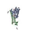
Mass: 24.305 Da / Num. of mol.: 1 / Source method: obtained synthetically / Formula: Mg
#4: Chemical ChemComp-GTP / GUANOSINE-5'-TRIPHOSPHATE / Guanosine triphosphate


Mass: 523.180 Da / Num. of mol.: 1 / Source method: obtained synthetically / Formula: C10H16N5O14P3 / Comment: GTP, energy-carrying molecule*YM
#5: Chemical ChemComp-ZN / ZINC ION


Mass: 65.409 Da / Num. of mol.: 2 / Source method: obtained synthetically / Formula: Zn
#6: Water ChemComp-HOH / water / Water


Mass: 18.015 Da / Num. of mol.: 46 / Source method: isolated from a natural source / Formula: H2O

-
Experimental details

-
Experiment

ExperimentMethod: X-RAY DIFFRACTION / Number of used crystals: 1

-
Sample preparation

CrystalDensity Matthews: 2.64 Å3/Da / Density % sol: 52.62 %
Crystal growpH: 8 / Details: pH 8.0
Crystal
*PLUS
Density % sol: 55 %
Crystal grow
*PLUS
Method: unknown
Components of the solutions
*PLUS
Conc.: 12.5 % / Common name: erythrotol

-
Data collection

DiffractionMean temperature: 100 K
Diffraction sourceSource: SYNCHROTRON / Site: NSLS / Beamline: X4A / Wavelength: 0.9789, 0.9793, 0.9879
DetectorType: RIGAKU / Detector: IMAGE PLATE / Date: May 31, 1998 / Details: MIRRORS
RadiationProtocol: MAD / Monochromatic (M) / Laue (L): M / Scattering type: x-ray
Radiation wavelength
IDWavelength (Å)Relative weight
10.97891
20.97931
30.98791
ReflectionResolution: 2.6→90 Å / Num. obs: 23889 / % possible obs: 98.5 % / Observed criterion σ(I): 0 / Redundancy: 2 % / Biso Wilson estimate: 35.1 Å2 / Rmerge(I) obs: 0.053 / Rsym value: 0.049 / Net I/σ(I): 17.9
Reflection shellResolution: 2.6→2.7 Å / Redundancy: 1.8 % / Rmerge(I) obs: 0.3 / Mean I/σ(I) obs: 0.3 / Rsym value: 0.22 / % possible all: 97.8
Reflection shell
*PLUS
% possible obs: 97.8 %

-
Processing

Software
NameVersionClassification
CNS0.4refinement
DENZOdata reduction
SCALEPACKdata scaling
CNS0.4phasing
RefinementMethod to determine structure: MAD / Resolution: 2.6→90 Å / Rfactor Rfree error: 0.006 / Data cutoff high rms absF: 174722 / Isotropic thermal model: RESTRAINED / Cross valid method: THROUGHOUT / σ(F): 0
RfactorNum. reflection% reflectionSelection details
Rfree0.263 2213 9.4 %RANDOM
Rwork0.226 ---
obs0.226 23478 96.8 %-
Solvent computationSolvent model: FLAT MODEL / Bsol: 49.72 Å2 / ksol: 0.33 e/Å3
Displacement parametersBiso mean: 51.5 Å2
Baniso -1Baniso -2Baniso -3
1-6.16 Å20 Å2-1.62 Å2
2--1.18 Å20 Å2
3----7.32 Å2
Refine analyze
FreeObs
Luzzati coordinate error0.43 Å0.34 Å
Luzzati d res low-5 Å
Luzzati sigma a0.44 Å0.35 Å
Refinement stepCycle: LAST / Resolution: 2.6→90 Å
ProteinNucleic acidLigandSolventTotal
Num. atoms2445 0 35 46 2526
Refine LS restraints
Refine-IDTypeDev idealDev ideal target
X-RAY DIFFRACTIONc_bond_d0.006
X-RAY DIFFRACTIONc_bond_d_na
X-RAY DIFFRACTIONc_bond_d_prot
X-RAY DIFFRACTIONc_angle_d
X-RAY DIFFRACTIONc_angle_d_na
X-RAY DIFFRACTIONc_angle_d_prot
X-RAY DIFFRACTIONc_angle_deg1.2
X-RAY DIFFRACTIONc_angle_deg_na
X-RAY DIFFRACTIONc_angle_deg_prot
X-RAY DIFFRACTIONc_dihedral_angle_d21.5
X-RAY DIFFRACTIONc_dihedral_angle_d_na
X-RAY DIFFRACTIONc_dihedral_angle_d_prot
X-RAY DIFFRACTIONc_improper_angle_d0.89
X-RAY DIFFRACTIONc_improper_angle_d_na
X-RAY DIFFRACTIONc_improper_angle_d_prot
X-RAY DIFFRACTIONc_mcbond_it6.521.5
X-RAY DIFFRACTIONc_mcangle_it8.82
X-RAY DIFFRACTIONc_scbond_it9.852
X-RAY DIFFRACTIONc_scangle_it12.392.5
LS refinement shellResolution: 2.6→2.76 Å / Rfactor Rfree error: 0.02 / Total num. of bins used: 6
RfactorNum. reflection% reflection
Rfree0.398 336 8.9 %
Rwork0.294 3456 -
obs--93.5 %
Xplor file
Refine-IDSerial noParam fileTopol file
X-RAY DIFFRACTION1PROTEIN_REP.PARAMPROTEIN.TOP
X-RAY DIFFRACTION2ION.PARAM
X-RAY DIFFRACTION3GTP.PAR
X-RAY DIFFRACTION4WATER.PARAM
Software
*PLUS
Name: CNS / Version: 0.4 / Classification: refinement
Refinement
*PLUS
Rfactor obs: 0.233 / Rfactor Rfree: 0.264
Solvent computation
*PLUS
Displacement parameters
*PLUS
Refine LS restraints
*PLUS
Refine-IDTypeDev ideal
X-RAY DIFFRACTIONc_dihedral_angle_d
X-RAY DIFFRACTIONc_dihedral_angle_deg21.5
X-RAY DIFFRACTIONc_improper_angle_d
X-RAY DIFFRACTIONc_improper_angle_deg0.89
LS refinement shell
*PLUS
Rfactor Rfree: 0.375 / Rfactor obs: 0.3

+
About Yorodumi

-
News

-
Feb 9, 2022. New format data for meta-information of EMDB entries

New format data for meta-information of EMDB entries

  • Version 3 of the EMDB header file is now the official format.
  • The previous official version 1.9 will be removed from the archive.

Related info.:EMDB header

External links:wwPDB to switch to version 3 of the EMDB data model

-
Aug 12, 2020. Covid-19 info

Covid-19 info

URL: https://pdbj.org/emnavi/covid19.php

New page: Covid-19 featured information page in EM Navigator.

Related info.:Covid-19 info / Mar 5, 2020. Novel coronavirus structure data

+
Mar 5, 2020. Novel coronavirus structure data

Novel coronavirus structure data

Related info.:Yorodumi Speices / Aug 12, 2020. Covid-19 info

External links:COVID-19 featured content - PDBj / Molecule of the Month (242):Coronavirus Proteases

+
Jan 31, 2019. EMDB accession codes are about to change! (news from PDBe EMDB page)

EMDB accession codes are about to change! (news from PDBe EMDB page)

  • The allocation of 4 digits for EMDB accession codes will soon come to an end. Whilst these codes will remain in use, new EMDB accession codes will include an additional digit and will expand incrementally as the available range of codes is exhausted. The current 4-digit format prefixed with “EMD-” (i.e. EMD-XXXX) will advance to a 5-digit format (i.e. EMD-XXXXX), and so on. It is currently estimated that the 4-digit codes will be depleted around Spring 2019, at which point the 5-digit format will come into force.
  • The EM Navigator/Yorodumi systems omit the EMD- prefix.

Related info.:Q: What is EMD? / ID/Accession-code notation in Yorodumi/EM Navigator

External links:EMDB Accession Codes are Changing Soon! / Contact to PDBj

+
Jul 12, 2017. Major update of PDB

Major update of PDB

  • wwPDB released updated PDB data conforming to the new PDBx/mmCIF dictionary.
  • This is a major update changing the version number from 4 to 5, and with Remediation, in which all the entries are updated.
  • In this update, many items about electron microscopy experimental information are reorganized (e.g. em_software).
  • Now, EM Navigator and Yorodumi are based on the updated data.

External links:wwPDB Remediation / Enriched Model Files Conforming to OneDep Data Standards Now Available in the PDB FTP Archive

-
Yorodumi

Thousand views of thousand structures

  • Yorodumi is a browser for structure data from EMDB, PDB, SASBDB, etc.
  • This page is also the successor to EM Navigator detail page, and also detail information page/front-end page for Omokage search.
  • The word "yorodu" (or yorozu) is an old Japanese word meaning "ten thousand". "mi" (miru) is to see.

Related info.:EMDB / PDB / SASBDB / Comparison of 3 databanks / Yorodumi Search / Aug 31, 2016. New EM Navigator & Yorodumi / Yorodumi Papers / Jmol/JSmol / Function and homology information / Changes in new EM Navigator and Yorodumi

Read more