 Movie
Movie Controller


+ Open data

- Basic information
| Entry | Database: PDB / ID: 4jmj | ||||||
|---|---|---|---|---|---|---|---|
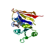
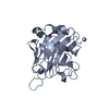
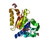
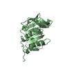
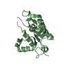
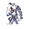
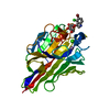
| Title | Structure of dusp11 | ||||||
 Components Components | RNA/RNP complex-1-interacting phosphatase | ||||||
 Keywords Keywords | HYDROLASE / alpha/beta hydrolase | ||||||
| Function / homology |  Function and homology information Function and homology informationpolynucleotide 5'-phosphatase activity / Hydrolases; Acting on ester bonds; Phosphoric-monoester hydrolases / protein dephosphorylation / phosphatase activity / RNA processing / intercellular bridge / protein tyrosine phosphatase activity / fibrillar center / nuclear speck / mitochondrion ...polynucleotide 5'-phosphatase activity / Hydrolases; Acting on ester bonds; Phosphoric-monoester hydrolases / protein dephosphorylation / phosphatase activity / RNA processing / intercellular bridge / protein tyrosine phosphatase activity / fibrillar center / nuclear speck / mitochondrion / RNA binding / nucleoplasm / nucleus Similarity search - Function | ||||||
| Biological species |  Homo sapiens (human) Homo sapiens (human) | ||||||
| Method |  X-RAY DIFFRACTION / SYNCHROTRON / MOLECULAR REPLACEMENT / Resolution: 2.382 Å X-RAY DIFFRACTION / SYNCHROTRON / MOLECULAR REPLACEMENT / Resolution: 2.382 Å | ||||||
 Authors Authors | Jeong, D.G. / Kim, S.J. / Ryu, S.E. | ||||||
 Citation Citation | Journal: Acta Crystallogr.,Sect.D / Year: 2014 Title: The family-wide structure and function of human dual-specificity protein phosphatases. Authors: Jeong, D.G. / Wei, C.H. / Ku, B. / Jeon, T.J. / Chien, P.N. / Kim, J.K. / Park, S.Y. / Hwang, H.S. / Ryu, S.Y. / Park, H. / Kim, D.S. / Kim, S.J. / Ryu, S.E. | ||||||
| History |
|
- Structure visualization
| Structure viewer | Molecule: MolmilJmol/JSmol |
|---|
- Downloads & links
-Download
| PDBx/mmCIF format | 4jmj.cif.gz | 89.9 KB | Display | PDBx/mmCIF format |
|---|---|---|---|---|
| PDB format | pdb4jmj.ent.gz | 68.3 KB | Display | PDB format |
| PDBx/mmJSON format | 4jmj.json.gz | Tree view | PDBx/mmJSON format | |
| Others |  Other downloads Other downloads |
-Validation report
| Arichive directory | https://data.pdbj.org/pub/pdb/validation_reports/jm/4jmjftp://data.pdbj.org/pub/pdb/validation_reports/jm/4jmj | HTTPS FTP |
|---|
-Related structure data
| Related structure data |  4jmkC 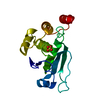 4jnbC 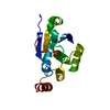 4ki9C  1yn9S C: citing same article ( S: Starting model for refinement |
|---|---|
| Similar structure data |


-Links
 PDBj
PDBj


- Assembly
| Deposited unit | 
| ||||||||
|---|---|---|---|---|---|---|---|---|---|
| 1 |
| ||||||||
| Unit cell |
|
-Components
| #1: Protein | Mass: 21309.303 Da / Num. of mol.: 1 / Fragment: UNP residues 28-208 / Mutation: C125S Source method: isolated from a genetically manipulated source Source: (gene. exp.) Homo sapiens (human) / Gene: DUSP11, PIR1 / Production host:  References: UniProt: O75319, Hydrolases; Acting on ester bonds; Phosphoric-monoester hydrolases |
|---|---|
| #2: Chemical | ChemComp-PO4 /   Mass: 94.971 Da / Num. of mol.: 1 / Source method: obtained synthetically / Formula: PO4 Mass: 94.971 Da / Num. of mol.: 1 / Source method: obtained synthetically / Formula: PO4 |
| #3: Chemical | ChemComp-CL /   Mass: 35.453 Da / Num. of mol.: 1 / Source method: obtained synthetically / Formula: Cl Mass: 35.453 Da / Num. of mol.: 1 / Source method: obtained synthetically / Formula: Cl |
| #4: Water | ChemComp-HOH /  Mass: 18.015 Da / Num. of mol.: 30 / Source method: isolated from a natural source / Formula: H2O Mass: 18.015 Da / Num. of mol.: 30 / Source method: isolated from a natural source / Formula: H2O |
-Experimental details
-Experiment
| Experiment | Method: X-RAY DIFFRACTION / Number of used crystals: 1 |
|---|
- Sample preparation
| Crystal | Density Matthews: 2.7 Å3/Da / Density % sol: 54.37 % |
|---|---|
| Crystal grow | Temperature: 291 K / Method: vapor diffusion, hanging drop / pH: 7 Details: PEG 4000, HEPES, pH 7.0, VAPOR DIFFUSION, HANGING DROP, temperature 291K |
-Data collection
| Diffraction | Mean temperature: 100 K |
|---|---|
| Diffraction source | Source: SYNCHROTRON / Site: PAL/PLS  / Beamline: 6B / Wavelength: 1 Å / Beamline: 6B / Wavelength: 1 Å |
| Detector | Type: ADSC QUANTUM 210 / Detector: CCD / Date: Oct 10, 2006 |
| Radiation | Monochromator: YALE MIRRORS / Protocol: SINGLE WAVELENGTH / Monochromatic (M) / Laue (L): M / Scattering type: x-ray |
| Radiation wavelength | Wavelength: 1 Å / Relative weight: 1 |
| Reflection | Resolution: 2.38→40 Å / Num. all: 9356 / Num. obs: 9276 / % possible obs: 99.2 % / Observed criterion σ(F): 0 / Observed criterion σ(I): 0 |
| Reflection shell | Resolution: 2.38→2.47 Å / % possible all: 94 |
- Processing
| Software |
| ||||||||||||||||||||||||||||||||||||||||
|---|---|---|---|---|---|---|---|---|---|---|---|---|---|---|---|---|---|---|---|---|---|---|---|---|---|---|---|---|---|---|---|---|---|---|---|---|---|---|---|---|---|
| Refinement | Method to determine structure: MOLECULAR REPLACEMENT Starting model: 1YN9 Resolution: 2.382→27.36 Å / Occupancy max: 1 / Occupancy min: 1 / SU ML: 0.26 / σ(F): 1.32 / Phase error: 26.28 / Stereochemistry target values: ML
| ||||||||||||||||||||||||||||||||||||||||
| Solvent computation | Shrinkage radii: 0.9 Å / VDW probe radii: 1.11 Å / Solvent model: FLAT BULK SOLVENT MODEL | ||||||||||||||||||||||||||||||||||||||||
| Displacement parameters | Biso max: 138.94 Å2 / Biso mean: 61.4401 Å2 / Biso min: 25.35 Å2 | ||||||||||||||||||||||||||||||||||||||||
| Refinement step | Cycle: LAST / Resolution: 2.382→27.36 Å
| ||||||||||||||||||||||||||||||||||||||||
| Refine LS restraints |
| ||||||||||||||||||||||||||||||||||||||||
| LS refinement shell | Refine-ID: X-RAY DIFFRACTION / Total num. of bins used: 4
| ||||||||||||||||||||||||||||||||||||||||
| Refinement TLS params. | Method: refined / Origin x: 35.9787 Å / Origin y: 11.8223 Å / Origin z: 3.0491 Å
| ||||||||||||||||||||||||||||||||||||||||
| Refinement TLS group | Selection details: all |
