 Movie
Movie Controller

[English] 日本語
 Yorodumi
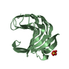
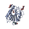
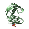
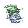
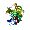
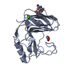
- PDB-6y0h: High resolution structure of GH11 xylanase from Nectria haematococca -
+ Open data

- Basic information
| Entry | Database: PDB / ID: 6y0h | ||||||
|---|---|---|---|---|---|---|---|
| Title | High resolution structure of GH11 xylanase from Nectria haematococca | ||||||
 Components Components | Endo-1,4-beta-xylanase | ||||||
 Keywords Keywords | HYDROLASE / Xylanase / Atomic resolution / Nectria haematococca | ||||||
| Function / homology |  Function and homology information Function and homology informationendo-1,4-beta-xylanase / endo-1,4-beta-xylanase activity / xylan catabolic process Similarity search - Function | ||||||
| Biological species |  [Nectria] haematococca mpVI 77-13-4 (fungus) [Nectria] haematococca mpVI 77-13-4 (fungus) | ||||||
| Method |  X-RAY DIFFRACTION / SYNCHROTRON / MOLECULAR REPLACEMENT / molecular replacement / Resolution: 1 Å X-RAY DIFFRACTION / SYNCHROTRON / MOLECULAR REPLACEMENT / molecular replacement / Resolution: 1 Å | ||||||
 Authors Authors | Andaleeb, H. / Betzel, C. / Perbandt, M. / Brognaro, H. | ||||||
 Citation Citation | Journal: Sci Rep / Year: 2020 Title: High-resolution crystal structure and biochemical characterization of a GH11 endoxylanase from Nectria haematococca. Authors: Andaleeb, H. / Ullah, N. / Falke, S. / Perbandt, M. / Brognaro, H. / Betzel, C. | ||||||
| History |
|
- Structure visualization
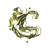
| Structure viewer | Molecule: MolmilJmol/JSmol |
|---|
- Downloads & links
-Download
| PDBx/mmCIF format | 6y0h.cif.gz | 101.6 KB | Display | PDBx/mmCIF format |
|---|---|---|---|---|
| PDB format | pdb6y0h.ent.gz | 77.5 KB | Display | PDB format |
| PDBx/mmJSON format | 6y0h.json.gz | Tree view | PDBx/mmJSON format | |
| Others |  Other downloads Other downloads |
-Validation report
| Arichive directory | https://data.pdbj.org/pub/pdb/validation_reports/y0/6y0hftp://data.pdbj.org/pub/pdb/validation_reports/y0/6y0h | HTTPS FTP |
|---|
-Related structure data
| Related structure data | 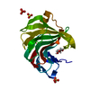 5jrmS S: Starting model for refinement |
|---|---|
| Similar structure data |


-Links
 PDBj
PDBj
- Assembly
| Deposited unit | 
| ||||||||
|---|---|---|---|---|---|---|---|---|---|
| 1 |
| ||||||||
| Unit cell |
| ||||||||
| Components on special symmetry positions |
|
-Components
| #1: Protein | Mass: 20917.430 Da / Num. of mol.: 1 Source method: isolated from a genetically manipulated source Details: endo-1,4-beta-xylanase activity Source: (gene. exp.) [Nectria] haematococca mpVI 77-13-4 (fungus)Gene: NECHADRAFT_106153 / Production host:  |
|---|---|
| #2: Water | ChemComp-HOH /  Mass: 18.015 Da / Num. of mol.: 277 / Source method: isolated from a natural source / Formula: H2O Mass: 18.015 Da / Num. of mol.: 277 / Source method: isolated from a natural source / Formula: H2O |
-Experimental details
-Experiment
| Experiment | Method: X-RAY DIFFRACTION / Number of used crystals: 1 |
|---|
- Sample preparation
| Crystal | Density Matthews: 2.32 Å3/Da / Density % sol: 47 % |
|---|---|
| Crystal grow | Temperature: 293.15 K / Method: vapor diffusion, hanging drop / pH: 5.5 / Details: 1 M Ammonium sulphate,100mM Sodium citrate |
-Data collection
| Diffraction | Mean temperature: 100 K / Serial crystal experiment: N |
|---|---|
| Diffraction source | Source: SYNCHROTRON / Site: PETRA III, EMBL c/o DESY  / Beamline: P13 (MX1) / Wavelength: 0.97 Å / Beamline: P13 (MX1) / Wavelength: 0.97 Å |
| Detector | Type: DECTRIS PILATUS 6M / Detector: PIXEL / Date: Dec 4, 2018 |
| Radiation | Protocol: SINGLE WAVELENGTH / Monochromatic (M) / Laue (L): M / Scattering type: x-ray |
| Radiation wavelength | Wavelength: 0.97 Å / Relative weight: 1 |
| Reflection | Resolution: 1→34.67 Å / Num. obs: 82307 / % possible obs: 93.3 % / Redundancy: 3.2 % / CC1/2: 0.99 / Net I/σ(I): 15.5 |
| Reflection shell | Resolution: 1→1.06 Å / Num. unique obs: 11904 / CC1/2: 0.85 |
-Phasing
| Phasing | Method: molecular replacement |
|---|
- Processing
| Software |
| |||||||||||||||||||||||||||||||||||||||||||||||||||||||||||||||||||||||||||||||||||||||||||||||||||||||||||||||||||||||||||||||||||||||||||||||||
|---|---|---|---|---|---|---|---|---|---|---|---|---|---|---|---|---|---|---|---|---|---|---|---|---|---|---|---|---|---|---|---|---|---|---|---|---|---|---|---|---|---|---|---|---|---|---|---|---|---|---|---|---|---|---|---|---|---|---|---|---|---|---|---|---|---|---|---|---|---|---|---|---|---|---|---|---|---|---|---|---|---|---|---|---|---|---|---|---|---|---|---|---|---|---|---|---|---|---|---|---|---|---|---|---|---|---|---|---|---|---|---|---|---|---|---|---|---|---|---|---|---|---|---|---|---|---|---|---|---|---|---|---|---|---|---|---|---|---|---|---|---|---|---|---|---|---|
| Refinement | Method to determine structure: MOLECULAR REPLACEMENT Starting model: 5JRM Resolution: 1→34.67 Å / Cor.coef. Fo:Fc: 0.976 / Cor.coef. Fo:Fc free: 0.973 / SU B: 0.897 / SU ML: 0.02 / Cross valid method: FREE R-VALUE / ESU R: 0.026 / ESU R Free: 0.025 Details: HYDROGENS HAVE BEEN ADDED IN THE RIDING POSITIONS U VALUES : REFINED INDIVIDUALLY
| |||||||||||||||||||||||||||||||||||||||||||||||||||||||||||||||||||||||||||||||||||||||||||||||||||||||||||||||||||||||||||||||||||||||||||||||||
| Solvent computation | Ion probe radii: 0.7 Å / Shrinkage radii: 0.7 Å / VDW probe radii: 1.1 Å | |||||||||||||||||||||||||||||||||||||||||||||||||||||||||||||||||||||||||||||||||||||||||||||||||||||||||||||||||||||||||||||||||||||||||||||||||
| Displacement parameters | Biso max: 63.58 Å2 / Biso mean: 9.386 Å2 / Biso min: 4.42 Å2
| |||||||||||||||||||||||||||||||||||||||||||||||||||||||||||||||||||||||||||||||||||||||||||||||||||||||||||||||||||||||||||||||||||||||||||||||||
| Refinement step | Cycle: final / Resolution: 1→34.67 Å
| |||||||||||||||||||||||||||||||||||||||||||||||||||||||||||||||||||||||||||||||||||||||||||||||||||||||||||||||||||||||||||||||||||||||||||||||||
| Refine LS restraints |
| |||||||||||||||||||||||||||||||||||||||||||||||||||||||||||||||||||||||||||||||||||||||||||||||||||||||||||||||||||||||||||||||||||||||||||||||||
| LS refinement shell | Resolution: 1→1.023 Å / Rfactor Rfree error: 0
|
