 Movie
Movie Controller


+ Open data

- Basic information
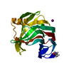
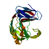
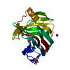
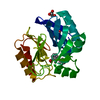
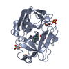
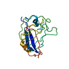
| Entry | Database: PDB / ID: 5zo0 | ||||||
|---|---|---|---|---|---|---|---|
| Title | Neutron structure of xylanase at pD5.4 | ||||||
 Components Components | Endo-1,4-beta-xylanase 2 | ||||||
 Keywords Keywords | HYDROLASE / family 11 GH / neutron crystallography / protonation state | ||||||
| Function / homology |  Function and homology information Function and homology informationendo-1,4-beta-xylanase / endo-1,4-beta-xylanase activity / xylan catabolic process / extracellular region Similarity search - Function | ||||||
| Biological species |  Hypocrea jecorina (fungus) Hypocrea jecorina (fungus) | ||||||
| Method | NEUTRON DIFFRACTION / NUCLEAR REACTOR /  MOLECULAR REPLACEMENT / Resolution: 1.648 Å MOLECULAR REPLACEMENT / Resolution: 1.648 Å | ||||||
 Authors Authors | Wan, Q. / Li, Z.H. | ||||||
| Funding support |  China, 1items China, 1items
| ||||||
 Citation Citation | #1: Journal: Proc. Natl. Acad. Sci. U.S.A. / Year: 2015Title: Direct determination of protonation states and visualization of hydrogen bonding in a glycoside hydrolase with neutron crystallography. Authors: Wan, Q. / Parks, J.M. / Hanson, B.L. / Fisher, S.Z. / Ostermann, A. / Schrader, T.E. / Graham, D.E. / Coates, L. / Langan, P. / Kovalevsky, A. #2: Journal: Acta Crystallogr. D Biol. Crystallogr. / Year: 2014Title: X-ray crystallographic studies of family 11 xylanase Michaelis and product complexes: implications for the catalytic mechanism. Authors: Wan, Q. / Zhang, Q. / Hamilton-Brehm, S. / Weiss, K. / Mustyakimov, M. / Coates, L. / Langan, P. / Graham, D. / Kovalevsky, A. | ||||||
| History |
|
- Structure visualization
| Structure viewer | Molecule: MolmilJmol/JSmol |
|---|
- Downloads & links
-Download
| PDBx/mmCIF format | 5zo0.cif.gz | 95.7 KB | Display | PDBx/mmCIF format |
|---|---|---|---|---|
| PDB format | pdb5zo0.ent.gz | 74.1 KB | Display | PDB format |
| PDBx/mmJSON format | 5zo0.json.gz | Tree view | PDBx/mmJSON format | |
| Others |  Other downloads Other downloads |
-Validation report
| Arichive directory | https://data.pdbj.org/pub/pdb/validation_reports/zo/5zo0ftp://data.pdbj.org/pub/pdb/validation_reports/zo/5zo0 | HTTPS FTP |
|---|
-Related structure data
| Related structure data |  2dfcS S: Starting model for refinement |
|---|---|
| Similar structure data | |
| Experimental dataset #1 | Data reference: 10.1073/pnas.1504986112 |


-Links
 PDBj
PDBj


- Assembly
| Deposited unit | 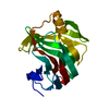
| ||||||||
|---|---|---|---|---|---|---|---|---|---|
| 1 |
| ||||||||
| Unit cell |
|
-Components
| #1: Protein | Mass: 20727.338 Da / Num. of mol.: 1 Source method: isolated from a genetically manipulated source Source: (gene. exp.) Hypocrea jecorina (strain ATCC 56765 / BCRC 32924 / NRRL 11460 / Rut C-30) (fungus)Strain: ATCC 56765 / BCRC 32924 / NRRL 11460 / Rut C-30 / Gene: xyn2, M419DRAFT_124931 / Plasmid: pET-NTMST / Details (production host): pET-28(b) derivative / Production host:  |
|---|---|
| #2: Chemical | ChemComp-DOD /   Mass: 18.015 Da / Num. of mol.: 154 / Source method: isolated from a natural source / Formula: D2O Mass: 18.015 Da / Num. of mol.: 154 / Source method: isolated from a natural source / Formula: D2O |
-Experimental details
-Experiment
| Experiment | Method: NEUTRON DIFFRACTION / Number of used crystals: 1 |
|---|
- Sample preparation
| Crystal | Density Matthews: 2.54 Å3/Da / Density % sol: 51.58 % |
|---|---|
| Crystal grow | Temperature: 295 K Details: The crystal was then sealed in a capillary with the D2O exchanged solution (2% PEG 3350, 0.2M NaI, 0.1M NaAc, pD5.4) for pH equilibration for two weeks before data collection (pD = pH + 0.4) Method: vapor diffusion, sitting drop / pH: 5 Details: 0.1M Tris, 0.1M NaCl, 1mM DTT, pH8.0, 2% PEG 3350, 0.2M NaI Temp details: room temperature |
-Data collection
| Diffraction | Mean temperature: 295 K | |||||||||||||||||||||||||||||||||||||||||||||||||||||||||||||||||||||||||||||||||||||||||||||||||||||||||||||||||||||
|---|---|---|---|---|---|---|---|---|---|---|---|---|---|---|---|---|---|---|---|---|---|---|---|---|---|---|---|---|---|---|---|---|---|---|---|---|---|---|---|---|---|---|---|---|---|---|---|---|---|---|---|---|---|---|---|---|---|---|---|---|---|---|---|---|---|---|---|---|---|---|---|---|---|---|---|---|---|---|---|---|---|---|---|---|---|---|---|---|---|---|---|---|---|---|---|---|---|---|---|---|---|---|---|---|---|---|---|---|---|---|---|---|---|---|---|---|---|---|
| Diffraction source | Source: NUCLEAR REACTOR / Site: FRM II  / Beamline: BIODIFF / Wavelength: 2.66 Å / Beamline: BIODIFF / Wavelength: 2.66 Å | |||||||||||||||||||||||||||||||||||||||||||||||||||||||||||||||||||||||||||||||||||||||||||||||||||||||||||||||||||||
| Detector | Type: BIODIFF / Detector: IMAGE PLATE / Date: Oct 1, 2017 | |||||||||||||||||||||||||||||||||||||||||||||||||||||||||||||||||||||||||||||||||||||||||||||||||||||||||||||||||||||
| Radiation | Monochromator: pyrolytic graphite / Protocol: SINGLE WAVELENGTH / Monochromatic (M) / Laue (L): M / Scattering type: neutron | |||||||||||||||||||||||||||||||||||||||||||||||||||||||||||||||||||||||||||||||||||||||||||||||||||||||||||||||||||||
| Radiation wavelength | Wavelength: 2.66 Å / Relative weight: 1 | |||||||||||||||||||||||||||||||||||||||||||||||||||||||||||||||||||||||||||||||||||||||||||||||||||||||||||||||||||||
| Reflection | Resolution: 1.65→50 Å / Num. obs: 22574 / % possible obs: 86.1 % / Redundancy: 2 % / Biso Wilson estimate: 7.29 Å2 / Rmerge(I) obs: 0.133 / Rpim(I) all: 0.103 / Rrim(I) all: 0.17 / Χ2: 0.991 / Net I/σ(I): 7.1 / Num. measured all: 44926 | |||||||||||||||||||||||||||||||||||||||||||||||||||||||||||||||||||||||||||||||||||||||||||||||||||||||||||||||||||||
| Reflection shell | Diffraction-ID: 1
|
- Processing
| Software |
| |||||||||||||||||||||||||||||||||||||||||||||||||||||||||||||||
|---|---|---|---|---|---|---|---|---|---|---|---|---|---|---|---|---|---|---|---|---|---|---|---|---|---|---|---|---|---|---|---|---|---|---|---|---|---|---|---|---|---|---|---|---|---|---|---|---|---|---|---|---|---|---|---|---|---|---|---|---|---|---|---|---|
| Refinement | Method to determine structure: MOLECULAR REPLACEMENT Starting model: 2dfc Resolution: 1.648→22.864 Å / SU ML: 0.23 / Cross valid method: THROUGHOUT / σ(F): 1.35 / Phase error: 22.03 / Stereochemistry target values: ML
| |||||||||||||||||||||||||||||||||||||||||||||||||||||||||||||||
| Solvent computation | Shrinkage radii: 0.9 Å / VDW probe radii: 1.11 Å / Solvent model: FLAT BULK SOLVENT MODEL | |||||||||||||||||||||||||||||||||||||||||||||||||||||||||||||||
| Displacement parameters | Biso max: 71.15 Å2 / Biso mean: 21.9251 Å2 / Biso min: 3.26 Å2 | |||||||||||||||||||||||||||||||||||||||||||||||||||||||||||||||
| Refinement step | Cycle: final / Resolution: 1.65→22.86 Å
| |||||||||||||||||||||||||||||||||||||||||||||||||||||||||||||||
| Refine LS restraints |
| |||||||||||||||||||||||||||||||||||||||||||||||||||||||||||||||
| LS refinement shell | Refine-ID: NEUTRON DIFFRACTION / Rfactor Rfree error: 0 / Total num. of bins used: 8
|
