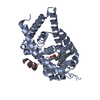
 Movie
Movie Controller


+ Open data

- Basic information
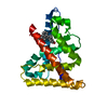
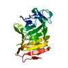
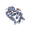
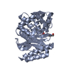
| Entry | Database: PDB / ID: 6xzv | |||||||||
|---|---|---|---|---|---|---|---|---|---|---|
| Title | Structure of zVDR LBD-Calcitriol in complex with chimera 18 | |||||||||
 Components Components |
| |||||||||
 Keywords Keywords | GENE REGULATION / Nuclear Receptor / Foldamer / Helix mimicry / Protein-protein Interaction | |||||||||
| Function / homology |  Function and homology information Function and homology informationheart jogging / Vitamin D (calciferol) metabolism / SUMOylation of intracellular receptors / calcitriol binding / lithocholic acid binding / vitamin D binding / heart looping / hematopoietic stem cell proliferation / calcium ion homeostasis / intracellular receptor signaling pathway ...heart jogging / Vitamin D (calciferol) metabolism / SUMOylation of intracellular receptors / calcitriol binding / lithocholic acid binding / vitamin D binding / heart looping / hematopoietic stem cell proliferation / calcium ion homeostasis / intracellular receptor signaling pathway / ossification / nuclear receptor activity / cell differentiation / RNA polymerase II cis-regulatory region sequence-specific DNA binding / regulation of DNA-templated transcription / DNA-templated transcription / negative regulation of transcription by RNA polymerase II / positive regulation of transcription by RNA polymerase II / zinc ion binding / nucleus / cytoplasm Similarity search - Function | |||||||||
| Biological species |  synthetic construct (others) | |||||||||
| Method |  X-RAY DIFFRACTION / SYNCHROTRON / MOLECULAR REPLACEMENT / Resolution: 2.3 Å X-RAY DIFFRACTION / SYNCHROTRON / MOLECULAR REPLACEMENT / Resolution: 2.3 Å | |||||||||
 Authors Authors | Buratto, J. / Belorusova, A.Y. / Rochel, N. / Guichard, G. | |||||||||
| Funding support |  France, 1items France, 1items
| |||||||||
 Citation Citation | Journal: Angew.Chem.Int.Ed.Engl. / Year: 2021 Title: Structural Basis for alpha-Helix Mimicry and Inhibition of Protein-Protein Interactions with Oligourea Foldamers. Authors: Cussol, L. / Mauran-Ambrosino, L. / Buratto, J. / Belorusova, A.Y. / Neuville, M. / Osz, J. / Fribourg, S. / Fremaux, J. / Dolain, C. / Goudreau, S.R. / Rochel, N. / Guichard, G. | |||||||||
| History |
|
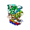
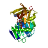
- Structure visualization
| Structure viewer | Molecule: MolmilJmol/JSmol |
|---|
- Downloads & links
-Download
| PDBx/mmCIF format | 6xzv.cif.gz | 120.9 KB | Display | PDBx/mmCIF format |
|---|---|---|---|---|
| PDB format | pdb6xzv.ent.gz | 89.8 KB | Display | PDB format |
| PDBx/mmJSON format | 6xzv.json.gz | Tree view | PDBx/mmJSON format | |
| Others |  Other downloads Other downloads |
-Validation report
| Arichive directory | https://data.pdbj.org/pub/pdb/validation_reports/xz/6xzvftp://data.pdbj.org/pub/pdb/validation_reports/xz/6xzv | HTTPS FTP |
|---|
-Related structure data
| Related structure data | 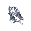 6hfaC 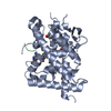 6xzhC  6xziC  6xzjC  6xzkC 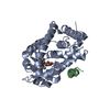 2hc4S S: Starting model for refinement C: citing same article ( |
|---|---|
| Similar structure data |


-Links
 PDBj
PDBj
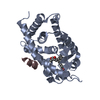
- Assembly
| Deposited unit | 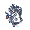
| ||||||||
|---|---|---|---|---|---|---|---|---|---|
| 1 |
| ||||||||
| Unit cell |
|
-Components
| #1: Protein | Mass: 34060.672 Da / Num. of mol.: 1 Source method: isolated from a genetically manipulated source Source: (gene. exp.)  |
|---|---|
| #2: Protein/peptide | Mass: 952.218 Da / Num. of mol.: 1 / Source method: obtained synthetically / Source: (synth.) synthetic construct (others) |
| #3: Chemical | ChemComp-VDX / 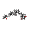  Mass: 416.636 Da / Num. of mol.: 1 / Source method: obtained synthetically / Formula: C27H44O3 Mass: 416.636 Da / Num. of mol.: 1 / Source method: obtained synthetically / Formula: C27H44O3 |
| #4: Water | ChemComp-HOH /  Mass: 18.015 Da / Num. of mol.: 38 / Source method: isolated from a natural source / Formula: H2O Mass: 18.015 Da / Num. of mol.: 38 / Source method: isolated from a natural source / Formula: H2O |
| Has ligand of interest | Y |
| Has protein modification | Y |
-Experimental details
-Experiment
| Experiment | Method: X-RAY DIFFRACTION / Number of used crystals: 1 |
|---|
- Sample preparation
| Crystal | Density Matthews: 2.27 Å3/Da / Density % sol: 45.88 % |
|---|---|
| Crystal grow | Temperature: 290 K / Method: vapor diffusion, hanging drop / Details: Lithium sulfate 1.5 M |
-Data collection
| Diffraction | Mean temperature: 100 K / Serial crystal experiment: N |
|---|---|
| Diffraction source | Source: SYNCHROTRON / Site: ESRF / Beamline: ID30B / Wavelength: 0.97 Å |
| Detector | Type: DECTRIS PILATUS 6M / Detector: PIXEL / Date: Sep 8, 2018 |
| Radiation | Protocol: SINGLE WAVELENGTH / Monochromatic (M) / Laue (L): M / Scattering type: x-ray |
| Radiation wavelength | Wavelength: 0.97 Å / Relative weight: 1 |
| Reflection | Resolution: 2.03→19.92 Å / Num. obs: 15432 / % possible obs: 99.34 % / Redundancy: 37.1 % / Rpim(I) all: 0.018 / Net I/σ(I): 25.56 |
| Reflection shell | Resolution: 2.03→2.38 Å / Redundancy: 40 % / Mean I/σ(I) obs: 2.06 / Num. unique obs: 1492 / Rpim(I) all: 0.372 / % possible all: 100 |
- Processing
| Software |
| |||||||||||||||||||||||||||||||||||||||||||||||||||||||||||||||||||||||||||||
|---|---|---|---|---|---|---|---|---|---|---|---|---|---|---|---|---|---|---|---|---|---|---|---|---|---|---|---|---|---|---|---|---|---|---|---|---|---|---|---|---|---|---|---|---|---|---|---|---|---|---|---|---|---|---|---|---|---|---|---|---|---|---|---|---|---|---|---|---|---|---|---|---|---|---|---|---|---|---|
| Refinement | Method to determine structure: MOLECULAR REPLACEMENT Starting model: 2HC4 Resolution: 2.3→19.92 Å / SU ML: 0.26 / Cross valid method: THROUGHOUT / σ(F): 1.35 / Phase error: 24.8 / Stereochemistry target values: ML
| |||||||||||||||||||||||||||||||||||||||||||||||||||||||||||||||||||||||||||||
| Solvent computation | Shrinkage radii: 0.9 Å / VDW probe radii: 1.11 Å / Solvent model: FLAT BULK SOLVENT MODEL | |||||||||||||||||||||||||||||||||||||||||||||||||||||||||||||||||||||||||||||
| Displacement parameters | Biso max: 156.88 Å2 / Biso mean: 74.0103 Å2 / Biso min: 43.48 Å2 | |||||||||||||||||||||||||||||||||||||||||||||||||||||||||||||||||||||||||||||
| Refinement step | Cycle: final / Resolution: 2.3→19.92 Å
| |||||||||||||||||||||||||||||||||||||||||||||||||||||||||||||||||||||||||||||
| Refine LS restraints |
| |||||||||||||||||||||||||||||||||||||||||||||||||||||||||||||||||||||||||||||
| LS refinement shell | Refine-ID: X-RAY DIFFRACTION / Rfactor Rfree error: 0 / Total num. of bins used: 10
| |||||||||||||||||||||||||||||||||||||||||||||||||||||||||||||||||||||||||||||
| Refinement TLS params. | Method: refined / Origin x: -31.6688 Å / Origin y: 25.9107 Å / Origin z: -1.6668 Å
| |||||||||||||||||||||||||||||||||||||||||||||||||||||||||||||||||||||||||||||
| Refinement TLS group |
|
