 Movie
Movie Controller

[English] 日本語
 Yorodumi
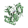
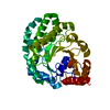
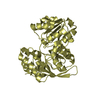
- PDB-6xyz: Crystal structure of the GH18 chitinase ChiB from the chitin util... -
+ Open data

- Basic information
| Entry | Database: PDB / ID: 6xyz | ||||||||||||
|---|---|---|---|---|---|---|---|---|---|---|---|---|---|
| Title | Crystal structure of the GH18 chitinase ChiB from the chitin utilization locus of Flavobacterium johnsoniae | ||||||||||||
 Components Components | Candidate chitinase Glycoside hydrolase family 18 | ||||||||||||
 Keywords Keywords | HYDROLASE / Chitinase GH18 Chitin ChiB | ||||||||||||
| Function / homology |  Function and homology information Function and homology informationchitin binding / hydrolase activity, hydrolyzing O-glycosyl compounds / carbohydrate metabolic process Similarity search - Function | ||||||||||||
| Biological species |  Flavobacterium johnsoniae (bacteria) Flavobacterium johnsoniae (bacteria) | ||||||||||||
| Method |  X-RAY DIFFRACTION / SYNCHROTRON / MOLECULAR REPLACEMENT / Resolution: 1.63 Å X-RAY DIFFRACTION / SYNCHROTRON / MOLECULAR REPLACEMENT / Resolution: 1.63 Å | ||||||||||||
 Authors Authors | Mazurkewich, S. / Helland, R. / MacKenzie, A. / Eijsink, V. / Pope, P. / Branden, G. / Larsbrink, J. | ||||||||||||
| Funding support |  Sweden, Sweden,  Norway, European Union, 3items Norway, European Union, 3items
| ||||||||||||
 Citation Citation | Journal: Sci Rep / Year: 2020 Title: Structural insights of the enzymes from the chitin utilization locus of Flavobacterium johnsoniae. Authors: Mazurkewich, S. / Helland, R. / Mackenzie, A. / Eijsink, V.G.H. / Pope, P.B. / Branden, G. / Larsbrink, J. | ||||||||||||
| History |
|
- Structure visualization
| Structure viewer | Molecule: MolmilJmol/JSmol |
|---|
- Downloads & links
-Download
| PDBx/mmCIF format | 6xyz.cif.gz | 95 KB | Display | PDBx/mmCIF format |
|---|---|---|---|---|
| PDB format | pdb6xyz.ent.gz | 63.8 KB | Display | PDB format |
| PDBx/mmJSON format | 6xyz.json.gz | Tree view | PDBx/mmJSON format | |
| Others |  Other downloads Other downloads |
-Validation report
| Arichive directory | https://data.pdbj.org/pub/pdb/validation_reports/xy/6xyzftp://data.pdbj.org/pub/pdb/validation_reports/xy/6xyz | HTTPS FTP |
|---|
-Related structure data
| Related structure data |  6yhhC  3fndS S: Starting model for refinement C: citing same article ( |
|---|---|
| Similar structure data |


-Links
 PDBj
PDBj


- Assembly
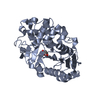
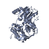
| Deposited unit | 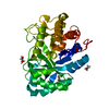
| ||||||||||||
|---|---|---|---|---|---|---|---|---|---|---|---|---|---|
| 1 |
| ||||||||||||
| Unit cell |
|
-Components
| #1: Protein | Mass: 36545.840 Da / Num. of mol.: 1 Source method: isolated from a genetically manipulated source Source: (gene. exp.) Flavobacterium johnsoniae (strain ATCC 17061 / DSM 2064 / UW101) (bacteria)Gene: Fjoh_4560 / Production host: | ||||||||
|---|---|---|---|---|---|---|---|---|---|
| #2: Chemical |   Mass: 62.068 Da / Num. of mol.: 3 / Source method: obtained synthetically / Formula: C2H6O2 Mass: 62.068 Da / Num. of mol.: 3 / Source method: obtained synthetically / Formula: C2H6O2#3: Chemical | ChemComp-FMT / |   Mass: 46.025 Da / Num. of mol.: 1 / Source method: obtained synthetically / Formula: CH2O2 Mass: 46.025 Da / Num. of mol.: 1 / Source method: obtained synthetically / Formula: CH2O2#4: Water | ChemComp-HOH / |  Mass: 18.015 Da / Num. of mol.: 338 / Source method: isolated from a natural source / Formula: H2O Mass: 18.015 Da / Num. of mol.: 338 / Source method: isolated from a natural source / Formula: H2OHas ligand of interest | N | Has protein modification | Y | |
-Experimental details
-Experiment
| Experiment | Method: X-RAY DIFFRACTION / Number of used crystals: 1 |
|---|
- Sample preparation
| Crystal | Density Matthews: 2.11 Å3/Da / Density % sol: 41.72 % |
|---|---|
| Crystal grow | Temperature: 295 K / Method: vapor diffusion, sitting drop / pH: 8 / Details: 0.15 M magnesium formate and 15% PEG3550 |
-Data collection
| Diffraction | Mean temperature: 100 K / Serial crystal experiment: N |
|---|---|
| Diffraction source | Source: SYNCHROTRON / Site: ESRF  / Beamline: MASSIF-3 / Wavelength: 0.9677 Å / Beamline: MASSIF-3 / Wavelength: 0.9677 Å |
| Detector | Type: DECTRIS EIGER X 4M / Detector: PIXEL / Date: May 3, 2017 |
| Radiation | Protocol: SINGLE WAVELENGTH / Monochromatic (M) / Laue (L): M / Scattering type: x-ray |
| Radiation wavelength | Wavelength: 0.9677 Å / Relative weight: 1 |
| Reflection | Resolution: 1.63→36.36 Å / Num. obs: 34484 / % possible obs: 90.73 % / Redundancy: 4.7 % / Biso Wilson estimate: 18.88 Å2 / CC1/2: 1 / Rmerge(I) obs: 0.04299 / Rpim(I) all: 0.02227 / Rrim(I) all: 0.04855 / Net I/σ(I): 19.71 |
| Reflection shell | Resolution: 1.63→1.688 Å / Redundancy: 4.8 % / Rmerge(I) obs: 0.2067 / Mean I/σ(I) obs: 6.25 / Num. unique obs: 2962 / CC1/2: 0.955 / Rpim(I) all: 0.1048 / Rrim(I) all: 0.2321 / % possible all: 77.85 |
- Processing
| Software |
| |||||||||||||||||||||||||||||||||||||||||||||||||||||||||||||||||||||||||||||||||||||||||||||||||||||||||
|---|---|---|---|---|---|---|---|---|---|---|---|---|---|---|---|---|---|---|---|---|---|---|---|---|---|---|---|---|---|---|---|---|---|---|---|---|---|---|---|---|---|---|---|---|---|---|---|---|---|---|---|---|---|---|---|---|---|---|---|---|---|---|---|---|---|---|---|---|---|---|---|---|---|---|---|---|---|---|---|---|---|---|---|---|---|---|---|---|---|---|---|---|---|---|---|---|---|---|---|---|---|---|---|---|---|---|
| Refinement | Method to determine structure: MOLECULAR REPLACEMENT Starting model: 3FND Resolution: 1.63→36.36 Å / SU ML: 0.1449 / Cross valid method: FREE R-VALUE / σ(F): 1.36 / Phase error: 20.8181 Stereochemistry target values: GeoStd + Monomer Library + CDL v1.2
| |||||||||||||||||||||||||||||||||||||||||||||||||||||||||||||||||||||||||||||||||||||||||||||||||||||||||
| Solvent computation | Shrinkage radii: 0.9 Å / VDW probe radii: 1.11 Å / Solvent model: FLAT BULK SOLVENT MODEL | |||||||||||||||||||||||||||||||||||||||||||||||||||||||||||||||||||||||||||||||||||||||||||||||||||||||||
| Displacement parameters | Biso mean: 24.59 Å2 | |||||||||||||||||||||||||||||||||||||||||||||||||||||||||||||||||||||||||||||||||||||||||||||||||||||||||
| Refinement step | Cycle: LAST / Resolution: 1.63→36.36 Å
| |||||||||||||||||||||||||||||||||||||||||||||||||||||||||||||||||||||||||||||||||||||||||||||||||||||||||
| Refine LS restraints |
| |||||||||||||||||||||||||||||||||||||||||||||||||||||||||||||||||||||||||||||||||||||||||||||||||||||||||
| LS refinement shell |
|
