 Movie
Movie Controller

[English] 日本語
 Yorodumi
- PDB-1jpi: Phe232Leu mutant of human UROD, human uroporphyrinogen III decarb... -
+ Open data

- Basic information
| Entry | Database: PDB / ID: 1jpi | ||||||
|---|---|---|---|---|---|---|---|
| Title | Phe232Leu mutant of human UROD, human uroporphyrinogen III decarboxylase | ||||||
 Components Components | UROPORPHYRINOGEN DECARBOXYLASE | ||||||
 Keywords Keywords | LYASE / heme biosynthesis | ||||||
| Function / homology |  Function and homology information Function and homology informationporphyrin-containing compound catabolic process / uroporphyrinogen decarboxylase / uroporphyrinogen decarboxylase activity / porphyrin-containing compound metabolic process / heme A biosynthetic process / heme B biosynthetic process / : / Heme biosynthesis / heme biosynthetic process / nucleoplasm / cytosol Similarity search - Function | ||||||
| Biological species |  Homo sapiens (human) Homo sapiens (human) | ||||||
| Method |  X-RAY DIFFRACTION / MOLECULAR REPLACEMENT / Resolution: 2.3 Å X-RAY DIFFRACTION / MOLECULAR REPLACEMENT / Resolution: 2.3 Å | ||||||
 Authors Authors | Phillips, J.D. / Parker, T.L. / Schubert, H.L. / Whitby, F.G. / Hill, C.P. / Kushner, J.P. | ||||||
 Citation Citation | Journal: Blood / Year: 2001 Title: Functional consequences of naturally occurring mutations in human uroporphyrinogen decarboxylase. Authors: Phillips, J.D. / Parker, T.L. / Schubert, H.L. / Whitby, F.G. / Hill, C.P. / Kushner, J.P. #1: Journal: Embo J. / Year: 1998Title: Crystal Structure of human uroporphyrinogen decarboxylase Authors: Whitby, F.G. / Phillips, J.D. / Kushner, J.P. / Hill, C.P. | ||||||
| History |
|
- Structure visualization
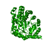
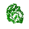
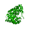
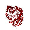
| Structure viewer | Molecule: MolmilJmol/JSmol |
|---|
- Downloads & links
-Download
| PDBx/mmCIF format | 1jpi.cif.gz | 89.5 KB | Display | PDBx/mmCIF format |
|---|---|---|---|---|
| PDB format | pdb1jpi.ent.gz | 66.5 KB | Display | PDB format |
| PDBx/mmJSON format | 1jpi.json.gz | Tree view | PDBx/mmJSON format | |
| Others |  Other downloads Other downloads |
-Validation report
| Arichive directory | https://data.pdbj.org/pub/pdb/validation_reports/jp/1jpiftp://data.pdbj.org/pub/pdb/validation_reports/jp/1jpi | HTTPS FTP |
|---|
-Related structure data
| Related structure data |  1jphC  1jpkC  1uroS S: Starting model for refinement C: citing same article ( |
|---|---|
| Similar structure data |


-Links
 PDBj
PDBj

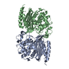
- Assembly
| Deposited unit | 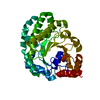
| ||||||||
|---|---|---|---|---|---|---|---|---|---|
| 1 | 
| ||||||||
| Unit cell |
| ||||||||
| Details | physiological dimer, monomer in the ASU |
-Components
| #1: Protein | Mass: 43331.477 Da / Num. of mol.: 1 / Mutation: F232L Source method: isolated from a genetically manipulated source Source: (gene. exp.) Homo sapiens (human) / Gene: UROD / Plasmid: pET14b / Production host:  |
|---|---|
| #2: Water | ChemComp-HOH /  Mass: 18.015 Da / Num. of mol.: 287 / Source method: isolated from a natural source / Formula: H2O Mass: 18.015 Da / Num. of mol.: 287 / Source method: isolated from a natural source / Formula: H2O |
-Experimental details
-Experiment
| Experiment | Method: X-RAY DIFFRACTION / Number of used crystals: 1 |
|---|
- Sample preparation
| Crystal | Density Matthews: 2.56 Å3/Da / Density % sol: 51.95 % | ||||||||||||||||||||||||||||||||||||||||||
|---|---|---|---|---|---|---|---|---|---|---|---|---|---|---|---|---|---|---|---|---|---|---|---|---|---|---|---|---|---|---|---|---|---|---|---|---|---|---|---|---|---|---|---|
| Crystal grow | Temperature: 298 K / Method: vapor diffusion, sitting drop / pH: 6.5 Details: MPD and MES, or Citrate, pH 6.5, VAPOR DIFFUSION, SITTING DROP, temperature 298K | ||||||||||||||||||||||||||||||||||||||||||
| Crystal grow | *PLUS Temperature: 21 ℃ / pH: 7.5 / Details: Phillips, J.D., (1997) Protein Sci., 6, 1343. | ||||||||||||||||||||||||||||||||||||||||||
| Components of the solutions | *PLUS
|
-Data collection
| Diffraction | Mean temperature: 100 K |
|---|---|
| Diffraction source | Source: ROTATING ANODE / Type: RIGAKU / Wavelength: 1.5418 |
| Detector | Type: RIGAKU RAXIS IV / Detector: IMAGE PLATE / Date: Jan 1, 1999 |
| Radiation | Monochromator: graphite / Protocol: SINGLE WAVELENGTH / Monochromatic (M) / Laue (L): M / Scattering type: x-ray |
| Radiation wavelength | Wavelength: 1.5418 Å / Relative weight: 1 |
| Reflection | Resolution: 2.3→20 Å / Num. obs: 19786 / % possible obs: 99 % / Observed criterion σ(F): 0 / Observed criterion σ(I): -3 / Rmerge(I) obs: 0.078 |
| Reflection shell | Resolution: 2.3→2.38 Å / Rmerge(I) obs: 0.34 / % possible all: 71.7 |
| Reflection | *PLUS Num. obs: 20018 / % possible obs: 99 % / Num. measured all: 262230 |
| Reflection shell | *PLUS Highest resolution: 2.3 Å / % possible obs: 99.7 % / Rmerge(I) obs: 0.4 |
- Processing
| Software |
| |||||||||||||||||||||||||||||||||||||||||||||||||
|---|---|---|---|---|---|---|---|---|---|---|---|---|---|---|---|---|---|---|---|---|---|---|---|---|---|---|---|---|---|---|---|---|---|---|---|---|---|---|---|---|---|---|---|---|---|---|---|---|---|---|
| Refinement | Method to determine structure: MOLECULAR REPLACEMENT Starting model: 1URO Resolution: 2.3→20 Å / Data cutoff high absF: 1000000 / Data cutoff low absF: 0.001 / σ(F): 0 / Stereochemistry target values: Engh and Huber
| |||||||||||||||||||||||||||||||||||||||||||||||||
| Refinement step | Cycle: LAST / Resolution: 2.3→20 Å
| |||||||||||||||||||||||||||||||||||||||||||||||||
| Refine LS restraints |
| |||||||||||||||||||||||||||||||||||||||||||||||||
| LS refinement shell |
| |||||||||||||||||||||||||||||||||||||||||||||||||
| Software | *PLUS Name: X-PLOR / Version: 3.843 / Classification: refinement | |||||||||||||||||||||||||||||||||||||||||||||||||
| Refinement | *PLUS Highest resolution: 2.3 Å / Lowest resolution: 20 Å / σ(F): 0 / % reflection Rfree: 5 % / Rfactor obs: 0.2 / Rfactor Rwork: 0.2 | |||||||||||||||||||||||||||||||||||||||||||||||||
| Solvent computation | *PLUS | |||||||||||||||||||||||||||||||||||||||||||||||||
| Displacement parameters | *PLUS | |||||||||||||||||||||||||||||||||||||||||||||||||
| Refine LS restraints | *PLUS Type: x_bond_d / Dev ideal: 0.14 | |||||||||||||||||||||||||||||||||||||||||||||||||
| LS refinement shell | *PLUS Highest resolution: 2.3 Å / Lowest resolution: 2.4 Å |
